


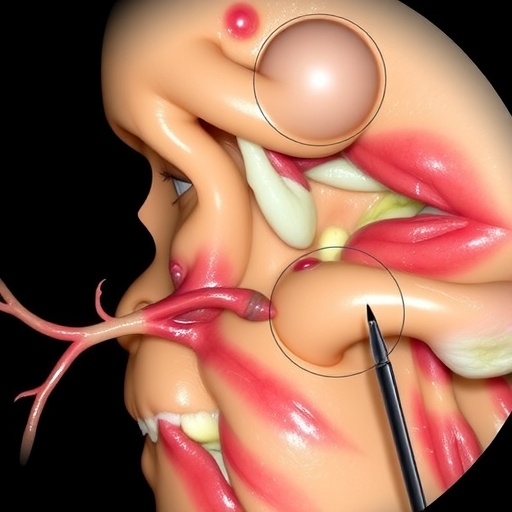
Vestibular schwannomas are benign tumors, but their location can make them among the most demanding growths to remove from the skull base. Arising from Schwann cells associated with the vestibular portion of the eighth cranial nerve, these tumors develop beside the brainstem and in the narrow cerebellopontine angle, where the facial nerve, cochlear nerve, major blood vessels and several other cranial nerves are tightly packed together. Surgeons treating them must therefore solve a difficult biological and technical problem: remove enough tumor to reduce the chance of regrowth while avoiding injury to the facial nerve, which controls expression, eye closure and many essential functions of the face. A newly reported retrospective study describes a microsurgical strategy designed to address both goals at once. Called subperineural “onion-peeling” dissection, the technique uses a thin membrane surrounding the tumor as a protective anatomical buffer while the tumor is separated from nearby nerves. In a series of 146 patients, the authors report that 95.2 percent eventually achieved normal or near-normal facial movement, while 97.3 percent underwent either gross-total or near-total tumor removal.
The study, published in the Journal of Neuro-Oncology, comes at a time when the traditional goal of gross-total resection is being reconsidered. Removing every visible fragment of a vestibular schwannoma can improve long-term tumor control, but aggressive dissection may place the facial nerve at risk, particularly when a large tumor has stretched or displaced it. To reduce that danger, many centers have adopted a planned subtotal resection followed by stereotactic radiosurgery. In this hybrid strategy, surgeons intentionally leave a portion of the tumor behind, then treat the remnant with focused radiation such as Gamma Knife or another stereotactic system. The approach can produce good facial-nerve outcomes, but it also means that patients may receive radiation even when the residual tumor might never grow. The authors of the new report argue that a more extensive microsurgical resection, when performed along the correct tissue plane, may preserve facial function without automatically committing patients to postoperative radiation.
The anatomical idea behind the operation is both simple and highly specialized. During tumor growth, the vestibular nerve fibers become stretched and attenuated around the mass. Their outer connective-tissue covering, described by the authors as the perineural membrane, may remain as a thin, translucent layer between the tumor and the facial and cochlear nerves. The surgeons compare this arrangement to the layers of an onion. The firm inner core represents the tumor itself, while the outer, more delicate layers correspond to the attenuated vestibular fibers and membrane. Instead of dissecting directly against the facial nerve, the surgeon develops a plane between the tumor parenchyma and the membrane. The facial nerve remains on the opposite, ventral side of the membrane, shielded from direct contact with surgical instruments. Intraoperatively, the membrane can appear almost transparent, leading the team to refer to it as a “holy veil.” Preserving it intact is intended to protect not only the facial nerve but also its small blood vessels and the exposed surface of the brainstem.
The operation begins with central debulking, in which the tumor’s interior is carefully hollowed out to create working space. Once the mass has been softened and reduced in volume, the surgeon can peel the remaining tumor away from the membrane under high magnification. The authors emphasize that the procedure is not a rigid attempt to remove every last cell at any cost. If the tumor separates cleanly, gross-total resection may be possible. If a portion is firmly attached to the membrane overlying the facial nerve, the surgeon sharply trims the tumor with microscissors and leaves only the smallest safe remnant. This result is classified as near-total resection when 95 to 99 percent of the tumor has been removed. A subtotal resection is defined as removal of less than 95 percent. The crucial difference from a pre-planned subtotal operation is that the decision to leave tumor is made during surgery, after the nerve’s position, tissue adherence and electrical responses have been assessed in real time.
In the 146-patient series, 89 patients underwent gross-total resection, 53 had near-total resection and four had subtotal resection. The group included a wide spectrum of tumor sizes, with diameters ranging from 0.3 to 6.4 centimeters and volumes ranging from 0.04 to 88.32 cubic centimeters. Nearly 70 percent were Koos grade III or IV tumors, meaning they were large enough to extend substantially into the cerebellopontine angle or compress the brainstem. Most operations were performed through a retrosigmoid approach, which accounted for 86.3 percent of cases; the remaining 13.7 percent used a translabyrinthine route. The facial nerve was continuously monitored with electromyography, and the team stimulated the nerve at the brainstem at the end of tumor removal. The mean stimulation threshold was 0.062 milliamperes, a low electrical current consistent with preserved nerve excitability.
Facial function was evaluated using the House-Brackmann grading system, in which grade I represents normal function and grade VI indicates complete paralysis. Before surgery, 98.6 percent of the patients were graded I and the remainder grade II. Immediately afterward, 84.2 percent still had grade I or II function. Temporary weakness was more common in the early postoperative period: 8.9 percent had grade III or IV function, while 6.8 percent had grade V or VI dysfunction. Over time, however, many of these patients improved. At the latest follow-up, 139 of the 146 patients, or 95.2 percent, had grade I or II facial function. Among those with immediate grade III or IV weakness, 12 of 13 recovered to grade I or II, typically within about six weeks. Recovery was slower and less complete among patients with the most severe early weakness, but some of these patients also returned to excellent function over several months.
The results suggest that a near-total resection did not necessarily produce worse facial outcomes than a gross-total resection. Among patients who underwent gross-total removal, 97.8 percent had grade I or II facial function at follow-up, compared with 92.5 percent after near-total resection. The difference was not statistically significant. Tumor size remained an important factor: all patients with Koos grade I or II tumors achieved favorable facial outcomes, compared with 93.1 percent of those with grade III or IV tumors. As tumors became larger, the proportion of gross-total resections declined and near-total resections increased. Gross-total removal was achieved in 93.75 percent of Koos I tumors, 86.2 percent of Koos II tumors, 76.9 percent of Koos III tumors and 39.5 percent of Koos IV tumors. The pattern reflects the surgeons’ willingness to leave a microscopic rim of tumor when attempting to remove it would place a displaced or stretched facial nerve under dangerous traction.
Longer-term tumor control was also encouraging, although the study was not large enough to establish definitive comparisons. During a mean follow-up of approximately 89.8 months, five patients experienced radiographic recurrence, producing an overall recurrence rate of 3.4 percent. Recurrence occurred in one of 89 gross-total resection cases, or 1.1 percent, and in four of 53 near-total resection cases, or 7.5 percent. The difference approached but did not reach conventional statistical significance. Three patients received stereotactic radiosurgery for recurrent or residual disease. Two remained stable after radiation, while one continued to progress and ultimately required another operation. Two other patients received radiosurgery for residual tumor that had not demonstrated progression and remained stable afterward. The postoperative cerebrospinal-fluid leak rate was 2.7 percent. Fourteen patients experienced some form of complication, and one patient died from intracranial hemorrhage after anticoagulation was administered for a postoperative heart attack.
The authors position the technique as an alternative to the increasingly popular planned subtotal resection followed by radiosurgery, but the comparison must be interpreted cautiously. The new study is retrospective, was conducted by a single surgeon at a high-volume skull-base center and did not directly randomize patients against a hybrid treatment group. Surgical expertise, patient selection, tumor anatomy and follow-up practices may therefore have influenced the results. The technique also has a steep learning curve. Identifying the perineural membrane requires detailed knowledge of the tumor-nerve interface, delicate microsurgical handling and the ability to change objectives during the operation. The surgeons describe stopping dissection when prolonged electrical activity, known as an A-train, appears during monitoring, irrigating the field and shifting to a safer subcapsular plane if necessary. These decisions depend heavily on experience and may not be reproducible immediately in lower-volume settings. In addition, hearing preservation was not the primary focus because many patients had large tumors and non-serviceable hearing before surgery.
Even with those limitations, the report highlights a broader shift in skull-base surgery: the most aggressive operation is not always the one that removes the greatest amount of tumor, and the safest operation is not necessarily the one that removes the least. The subperineural approach attempts to redefine that balance by treating the membrane around the vestibular nerve as a surgical guide and a biological shield. Its reported outcomes suggest that surgeons may be able to pursue gross-total or near-total resection while retaining the option of stopping when the facial nerve becomes vulnerable. Longer follow-up, independent replication and multicenter studies will be needed to determine whether the low recurrence rate persists for decades and whether other surgeons can achieve comparable facial-nerve results. For now, the “onion-peeling” concept offers a technically demanding but potentially powerful example of how microscopic anatomy can transform a high-risk operation into a more carefully calibrated contest between tumor control and neurological preservation.
Cite Scienmag News
Ophelia Keating. (August 26, 2026). Onion-Peeling Dissection Balances Resection and Facial Nerve Preservation in Vestibular Schwannoma Surgery. Scienmag. https://scienmag.com/onion-peeling-dissection-balances-resection-and-facial-nerve-preservation-in-vestibular-schwannoma-surgery/
Ophelia Keating. "Onion-Peeling Dissection Balances Resection and Facial Nerve Preservation in Vestibular Schwannoma Surgery." Scienmag, 26 August 2026, https://scienmag.com/onion-peeling-dissection-balances-resection-and-facial-nerve-preservation-in-vestibular-schwannoma-surgery/. Accessed 3 September 2026.
Ophelia Keating. "Onion-Peeling Dissection Balances Resection and Facial Nerve Preservation in Vestibular Schwannoma Surgery." Scienmag. August 26, 2026. https://scienmag.com/onion-peeling-dissection-balances-resection-and-facial-nerve-preservation-in-vestibular-schwannoma-surgery/


