


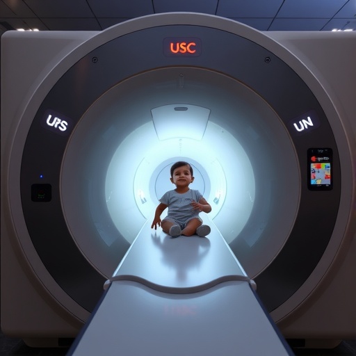
Some of the smallest patients in medicine create some of the biggest challenges for magnetic resonance imaging. An infant’s heart may be no larger than a walnut, yet it can beat at more than twice the rate of an adult heart while the child’s body changes rapidly with growth. Conventional MRI equipment, by contrast, is generally produced in standardized sizes designed around adult anatomy. Custom coils can improve the fit, but they often cost thousands of dollars and may require months or even years to manufacture. A team of engineers, imaging scientists and clinicians at the University of Southern California has now developed a radically faster and cheaper alternative: flexible MRI coils that can be customized digitally and 3D-printed in less than 10 minutes for approximately $30.
In tests, the experimental sensors generated about four times greater image contrast than standard commercial coils, a performance difference that could be especially important when physicians need to visualize small, moving structures. The technology was developed by researchers led by Yasser Khan, an assistant professor of electrical and computer engineering and biomedical engineering at the USC Viterbi School of Engineering. Their work, published in Nature Communications, demonstrates that flexible, patient-specific coils can improve imaging at a 0.55-tesla MRI field strength while also enabling dynamic scans of anatomy such as the wrist and heart. The researchers say the approach could eventually make advanced imaging more accessible to infants, children and other patients whose bodies do not conform well to the rigid dimensions of conventional equipment.
MRI scanners create images by placing the body inside a powerful magnetic field and using radiofrequency waves to disturb hydrogen nuclei, most commonly the protons in water molecules. As those protons return to their original state, they emit faint signals that are detected by radiofrequency coils and mathematically reconstructed into images. The closer and more precisely a coil fits around the anatomy being examined, the more efficiently it can capture those signals. A coil that is too large leaves gaps between the sensor and the body, reducing sensitivity and allowing noise to compete with the desired signal. For a small child, an adult-sized coil can therefore function like a large camera lens aimed at a tiny object: it may record the subject, but not with the sharpness or contrast that a properly matched lens could provide.
Khan’s team designed its sensors to follow the contours of the body rather than forcing patients into the fixed geometry of a rigid device. The coils are produced by printing conductive silver ink onto a thermoplastic elastomer, a soft polymer with mechanical properties similar to those of human skin. The resulting structure can stretch by roughly 5% to 10% and bend as the body moves, helping maintain close contact during imaging. That flexibility is more than a comfort feature. It can preserve the electrical relationship between the coil and the anatomy as a patient breathes, shifts position or moves a joint, reducing the loss of sensitivity that can occur when a rigid detector separates from the skin.
Creating the coils required the researchers to solve several engineering problems at once. They tested different flexible materials, plastics and printable metal formulations before selecting a combination that could provide both mechanical compliance and the electrical conductivity needed for MRI signal detection. They also developed the electronic interfaces required to connect the unconventional printed sensors to an MRI system. Unlike conventional coils, which are assembled from fixed components and manufactured through established industrial processes, the new devices begin as digital designs. Their dimensions, curvature and layout can be changed on a computer, allowing a new version to be printed almost immediately for a different patient, body region or imaging protocol.
That digital workflow could be particularly valuable in pediatric medicine, where a device that fits a child today may no longer be appropriate after months of growth. Instead of waiting for a replacement coil to be designed, fabricated and shipped, clinicians could theoretically create a new model based on updated measurements or a three-dimensional scan of the patient. The low material cost also changes the economics of customization. A coil that costs about $30 in materials and can be printed in minutes may be practical for repeated resizing, whereas a conventionally manufactured custom component costing thousands of dollars is far more difficult to replace or modify. The technology is not presented as a complete substitute for the safety testing and regulatory validation required before clinical deployment, but it offers a route toward equipment that can evolve with the patient.
The USC researchers are also targeting one of MRI’s most technically demanding applications: imaging anatomy in motion. Conventional MRI is often associated with still images because movement can blur the data and make reconstruction more difficult. Yet many important clinical questions concern structures that never stop moving, including the heart, lungs, joints and the developing fetal cardiovascular system. The new coils were tested with specialized MRI technology at the USC Michelson Center for Convergent Bioscience and the Dynamic Imaging Science Center, or DISC. The facility includes an unusual MRI system that researchers describe as the only one of its kind, designed to help capture the body while it moves and to support experiments in real-time imaging.
The collaboration brought together Khan’s expertise in flexible and wearable electronics with the MRI engineering capabilities of Krishna Shrinivas Nayak, a professor of electrical and computer engineering and biomedical engineering at USC Viterbi. Clinical insight came from John Wood, director of cardiovascular MRI at Children’s Hospital Los Angeles and a professor of pediatrics and radiology at the Keck School of Medicine of USC. Wood works with Nayak on difficult pediatric imaging problems, including real-time visualization of the fetal heart. Such examinations require sensors that can collect strong signals from small structures while accommodating rapid motion, changing anatomy and the practical difficulties of imaging patients who may be unable to remain completely still.
The reported improvement in image contrast is important because contrast determines how clearly tissues and structures can be distinguished from their surroundings. Higher contrast can help reveal boundaries that might otherwise be obscured by noise or weak signal, potentially giving clinicians more information without simply increasing the scan time or magnetic field strength. The study focused on imaging the wrist and heart at 0.55 tesla, a field strength lower than that used in many hospital MRI systems. Demonstrating useful performance under these conditions suggests that flexible, closely fitted coils could help compensate for some limitations of lower-field imaging, which is attracting interest because such scanners may be less expensive, more accessible and easier to deploy in settings where high-field systems are unavailable.
The researchers emphasize that the advance is not only a new medical device but also a new way of manufacturing MRI hardware. By combining printable electronics, compliant materials and rapid digital design, the system could allow imaging equipment to be tailored to anatomy rather than requiring anatomy to fit a limited set of standardized accessories. For infants with congenital heart disease, children undergoing repeated examinations and patients whose anatomy is difficult to accommodate, that shift could mean clearer images and more adaptable care. The next steps will involve further testing, refinement of the electronics and evaluation of how the coils perform across different patients, body regions and clinical conditions. If those studies confirm the early results, a low-cost sensor printed in minutes could help transform MRI from a largely standardized technology into one capable of adapting to the person inside the scanner.
Web References: https://viterbischool.usc.edu/news/2026/05/usc-researchers-develop-3d-printable-mri-coils-for-low-cost-improved-dynamic-imaging/; https://michelson.usc.edu/; https://disc.usc.edu/; https://khan.usc.edu/
References: Nature Communications, DOI: 10.1038/s41467-026-71817-x
Cite Scienmag News
Ophelia Keating. (August 25, 2026). New USC technology delivers clearer MRI scans for the tiniest patients. Scienmag. https://scienmag.com/new-usc-technology-delivers-clearer-mri-scans-for-the-tiniest-patients/
Ophelia Keating. "New USC technology delivers clearer MRI scans for the tiniest patients." Scienmag, 25 August 2026, https://scienmag.com/new-usc-technology-delivers-clearer-mri-scans-for-the-tiniest-patients/. Accessed 3 September 2026.
Ophelia Keating. "New USC technology delivers clearer MRI scans for the tiniest patients." Scienmag. August 25, 2026. https://scienmag.com/new-usc-technology-delivers-clearer-mri-scans-for-the-tiniest-patients/


