


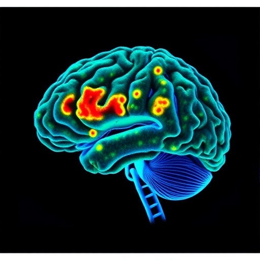
Alzheimer’s disease may be leaving a more precise anatomical signature in the brain than scientists once realized. A new study published in Translational Psychiatry investigates regional changes in the fine structure of brain tissue immediately beneath the cerebral cortex, known as the juxtacortical region, and links those changes to patterns of gene expression associated with neurodegeneration. The work, led by J. Zhang, L. Sun, X. Yang and colleagues, offers a combined view of Alzheimer’s biology that connects what can be detected through brain imaging with the molecular programs active inside vulnerable neural tissue.
The juxtacortical zone is a narrow but biologically important interface. It lies directly beneath the cortex, the outer layer responsible for functions including memory, language, perception and decision-making. Although much Alzheimer’s research has focused on cortical thinning, large-scale tissue loss and abnormalities in deeper brain structures, the tissue bordering the cortex may reveal earlier or more regionally specific changes. Its organization includes densely packed axons, dendrites, neurons, glial cells and blood vessels, all of which can be affected as the disease progresses. Subtle disruption in this area could therefore provide clues before dramatic anatomical damage becomes visible.
The study’s central idea is that Alzheimer’s disease does not alter every brain region in the same way. Instead, neurodegeneration follows a complex geography shaped by cellular composition, connectivity, metabolism and genetic vulnerability. By examining regional juxtacortical microstructural changes, the researchers seek to move beyond a simple question of whether tissue has shrunk. Microstructure refers to the organization of brain components at scales smaller than conventional anatomical imaging can directly resolve. Variations in water diffusion, tissue organization and local architecture can serve as indirect markers of changes to axons, membranes, extracellular space and other microscopic features.
This regional perspective matters because the same imaging signal can arise from different biological processes. A change in tissue organization may reflect loss of neuronal connections, damage to myelinated fibers, altered glial activity, inflammation or shifts in the balance of cellular compartments. Imaging alone can reveal where abnormalities are concentrated, but it cannot always explain why they occur. The researchers therefore paired their investigation of juxtacortical structure with gene-expression information, creating a bridge between regional anatomy and molecular biology. That combination can help identify which biological pathways may underlie the imaging patterns seen in Alzheimer’s disease.
Gene expression describes the activity of genes within cells and tissues: which genetic instructions are being read, and at what levels. In the Alzheimer’s brain, these molecular signatures can point toward processes such as synaptic dysfunction, immune activation, impaired energy production, protein handling and cellular stress. Mapping gene-expression patterns onto regions showing microstructural alterations may reveal whether vulnerable areas share particular molecular characteristics. It may also help distinguish tissue changes related directly to neurodegeneration from those associated with aging or general brain atrophy. The result is a more layered portrait of disease, linking microscopic organization to the biological machinery that maintains it.
The findings described by the paper’s title suggest that these two forms of evidence—regional microstructure and gene activity—provide complementary insights into Alzheimer’s pathology. Rather than treating the brain as a uniform organ undergoing gradual damage, the research emphasizes a mosaic of vulnerability. Some areas may show distinctive structural disruption alongside gene-expression profiles associated with neuronal injury or altered support-cell function, while other regions may be relatively resilient. Such differences could help explain why symptoms vary between individuals and why memory, language, attention and other abilities can deteriorate along different trajectories.
The work could also influence how scientists think about biomarkers. A biomarker is a measurable biological feature that helps indicate disease presence, progression or response to treatment. Traditional markers of Alzheimer’s include abnormal amyloid and tau proteins, brain volume loss and changes in metabolism. Juxtacortical microstructural measures, especially when interpreted alongside molecular data, could eventually add regional detail to this toolkit. They might help researchers identify tissue at risk, track the spread of degeneration or evaluate whether an experimental therapy is protecting neural architecture even before clinical improvement becomes obvious.
However, the study should not be interpreted as proof that a single imaging signature can diagnose Alzheimer’s disease or predict an individual’s future with certainty. Microstructural measurements are indirect, and gene-expression data often come from specialized datasets that may not capture every cell type or every stage of disease. Differences in age, disease severity, medication, vascular health and individual brain organization can also influence results. The strength of the approach lies not in one isolated measurement, but in the convergence of regional imaging and molecular evidence. Together, these methods can generate testable hypotheses about how and where neurodegeneration unfolds.
The broader message is that Alzheimer’s research is moving toward increasingly integrated maps of the human brain. The study by Zhang, Sun, Yang and colleagues presents the juxtacortical region as a potentially informative frontier, where microscopic tissue organization and gene regulation intersect. If future research confirms these relationships in larger and more diverse groups, the approach could sharpen disease staging and improve the design of targeted therapies. For now, the findings add weight to a crucial principle in neuroscience: understanding Alzheimer’s requires knowing not only which brain regions are damaged, but also how their cellular architecture and molecular identity make them vulnerable.
Cite Scienmag News
Cassandra Pierce. (August 2, 2026). Brain-region microstructure and gene activity reveal Alzheimer’s neurodegeneration. Scienmag. https://scienmag.com/brain-region-microstructure-and-gene-activity-reveal-alzheimers-neurodegeneration/
Cassandra Pierce. "Brain-region microstructure and gene activity reveal Alzheimer’s neurodegeneration." Scienmag, 2 August 2026, https://scienmag.com/brain-region-microstructure-and-gene-activity-reveal-alzheimers-neurodegeneration/. Accessed 4 September 2026.
Cassandra Pierce. "Brain-region microstructure and gene activity reveal Alzheimer’s neurodegeneration." Scienmag. August 2, 2026. https://scienmag.com/brain-region-microstructure-and-gene-activity-reveal-alzheimers-neurodegeneration/


