


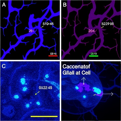
In a study published this year in Translational Psychiatry, researchers report that brain-resident vascular and glial cells can mount a coordinated response during sepsis—an immune-driven condition that can rapidly destabilize the nervous system. Using a pig model designed to mimic clinically relevant sepsis physiology, the team focused on how cells lining blood vessels and supporting glia communicate when systemic inflammation reaches the brain.
The investigators examined how vascular cell programs and glial signaling evolve under septic stress. Rather than treating brain inflammation as a set of independent events, the work emphasizes temporal alignment: molecular changes in the neurovascular unit appear to unfold in a coordinated manner, suggesting intercellular control across cell types. This coordination may help explain why some septic patients develop persistent cognitive and neurological impairments.
Technically, the study centers on mapping cell-state changes that reflect vascular activation alongside glial reactivity. Sepsis is known to disrupt endothelial function, compromise the blood–brain barrier, and alter inflammatory tone. Here, the authors argue that glia do not merely respond downstream; instead, they appear to participate in shaping vascular behavior, potentially stabilizing or amplifying inflammatory signaling pathways.
The pig platform is a key feature. Compared with smaller rodent models, large-animal physiology can better approximate human neurovascular dynamics. That translational relevance strengthens the case for targeting mechanisms that influence both blood vessels and glia simultaneously, rather than addressing them separately.
Importantly, the findings highlight the concept of a unified neuroimmune response. In sepsis, cytokines and danger signals circulate system-wide. The brain then integrates those cues through neurovascular coupling, where glial cells can influence local vascular signaling, permeability-related pathways, and inflammatory recruitment.
By characterizing coordinated cellular responses, the work also provides a framework for identifying biomarkers that span multiple cell compartments. Such biomarkers could help distinguish septic states that are more likely to progress to encephalopathy from those that resolve with standard care.
The study’s translational direction is clear: if coordination between vascular and glial programs can be modulated, new therapies may reduce brain injury during sepsis. Future experiments will likely test whether disrupting specific signaling axes can improve neurological outcomes without compromising systemic immune control.
Overall, the results place cell communication at the center of sepsis-associated brain dysfunction, offering a more integrated view of how the neurovascular unit behaves when the body’s inflammatory balance collapses.
Cite Scienmag News
Cassandra Pierce. (July 26, 2026). Coordinated Vascular and Glial Cell Response in Pig Sepsis Model. Scienmag. https://scienmag.com/coordinated-vascular-and-glial-cell-response-in-pig-sepsis-model/
Cassandra Pierce. "Coordinated Vascular and Glial Cell Response in Pig Sepsis Model." Scienmag, 26 July 2026, https://scienmag.com/coordinated-vascular-and-glial-cell-response-in-pig-sepsis-model/. Accessed 4 September 2026.
Cassandra Pierce. "Coordinated Vascular and Glial Cell Response in Pig Sepsis Model." Scienmag. July 26, 2026. https://scienmag.com/coordinated-vascular-and-glial-cell-response-in-pig-sepsis-model/


