


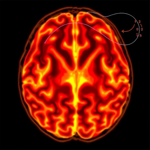
Scientists are turning photoacoustic computed tomography (PACT) into a more powerful tool for mapping the brain’s microvasculature and oxygen status by redesigning the ultrasound detection hardware. The approach targets a major bottleneck in conventional PACT: piezoelectric transducers limit how much depth resolution and functional detail can be captured in whole-brain imaging.
PACT works by converting absorbed optical energy into ultrasound, letting researchers visualize blood vessels with strong optical contrast and probe deeper than purely optical techniques. Yet deep imaging has been constrained by the need to balance sensitivity and spatial resolution using transducers tailored to different frequency bands—often requiring multiple detectors and complex alignment.
A new study in Light: Science & Applications introduces a sheet-like, dual-frequency fiber ultrasound transducer (FUT) concept for mouse brain imaging at the full-brain scale. The system uses a curved array of eight optical-fiber laser cavities, engineered so ultrasound focusing occurs at the geometrical center plane, enabling multispectral PACT without frequency-hopping hardware.
At the device level, the key innovation is how energy is redistributed between high- and low-frequency acoustic bands. By exploiting tight mechanical coupling between a quartz optical fiber and a polymer coating, the transducer transfers part of the fiber’s intrinsic high-frequency vibrational energy into a lower-frequency range. As a result, one element can simultaneously acquire dual-frequency photoacoustic signals, improving deep-tissue detection while preserving fine vascular sensitivity.
A second advance is the lens-less sheet-focused geometry. Both the bare silica fiber and the polymer coating are uniformly bent into an arc with a 4 cm curvature radius, naturally generating a sheet-like ultrasound focus. This eliminates bulky acoustic lenses and helps maintain accurate dual-frequency confocal alignment, reducing alignment and co-registration complexity.
Performance highlights include an ultra-low detection limit of approximately 5.2 Pa at a 4 cm working distance, a thin focus layer with ~400 µm slice thickness that suppresses out-of-plane interference, and confocal dual-frequency alignment that simplifies image formation.
The authors emphasize that the energy split between frequency bands can be tuned by adjusting the polymer-to-fiber length ratio, allowing the response to be customized for different imaging scenarios. Looking forward, scaling up FUT array element counts could remove mechanical scanning and support real-time whole-brain imaging and metabolism assessment.
This dual-frequency FUT array-based PACT strategy promises to overcome fundamental constraints of conventional ultrasound hardware, potentially accelerating both basic neuroscience studies and future clinical translation for brain disease diagnostics.
Keywords
Dual-frequency, fiber ultrasound transducer, FUT array, photoacoustic computed tomography, brain oxygen saturation, deep-tissue imaging, sheet-like focusing, high-resolution vascular imaging, optical-acoustic hybrid imaging
Cite Scienmag News
Cassandra Pierce. (July 14, 2026). Dual-Frequency Photoacoustic CT Reveals Clear Whole Brain, Centimeters Deep. Scienmag. https://scienmag.com/dual-frequency-photoacoustic-ct-reveals-clear-whole-brain-centimeters-deep/
Cassandra Pierce. "Dual-Frequency Photoacoustic CT Reveals Clear Whole Brain, Centimeters Deep." Scienmag, 14 July 2026, https://scienmag.com/dual-frequency-photoacoustic-ct-reveals-clear-whole-brain-centimeters-deep/. Accessed 4 September 2026.
Cassandra Pierce. "Dual-Frequency Photoacoustic CT Reveals Clear Whole Brain, Centimeters Deep." Scienmag. July 14, 2026. https://scienmag.com/dual-frequency-photoacoustic-ct-reveals-clear-whole-brain-centimeters-deep/


