In a groundbreaking study that bridges cellular biology and exercise physiology, researchers have unlocked a molecular mechanism that elucidates how physical activity drives bone formation, highlighting the pivotal role of primary cilia on preosteoclasts. This discovery, recently published in Experimental & Molecular Medicine, paves the way for novel therapeutic targets to enhance skeletal health and counteract osteoporosis and other bone-degenerative conditions. The research elucidates how exercise-induced mechanical stimuli translate into cellular signals, orchestrating periosteal bone formation through a previously underappreciated organelle — the primary cilium.
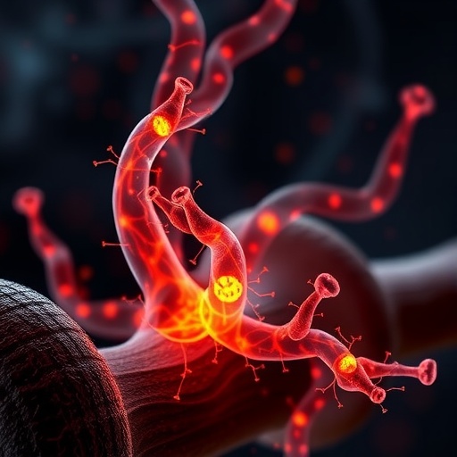
The primary cilium, a solitary, antenna-like projection found on nearly every mammalian cell, has emerged as a crucial sensory organelle responsible for detecting extracellular mechanical and chemical signals. In the context of bone biology, the functionality of primary cilia has remained enigmatic, particularly in preosteoclasts, the precursor cells to osteoclasts responsible for bone resorption. This new study presents compelling evidence that exercise triggers mechanosensitive responses in these cells via their primary cilia, promoting a local microenvironment conducive to periosteal bone accretion.
Delving deeper into the molecular cascade, the researchers demonstrated that mechanical loading from exercise stimulates the elongation and activation of primary cilia on preosteoclasts. This morphological transformation enhances the cells’ ability to sense biomechanical cues, triggering intracellular signaling pathways that modulate gene expression relevant to bone remodeling. Notably, the activation of these cilia leads to the secretion of osteogenic factors that directly facilitate periosteal bone formation, suggesting an autocrine and paracrine communication network within the bone niche.
This study employed advanced imaging techniques, including high-resolution confocal microscopy and live-cell imaging, to visualize primary cilium dynamics in response to mechanical stimuli. The dynamically elongated cilia serve as mechanotransducers, converting biomechanical inputs into biochemical signals that engage downstream effectors, including calcium influx channels and Hedgehog signaling pathways. Such mechanotransduction underscores the intimate relationship between physical activity and bone homeostasis, providing a refined understanding of how mechanical forces optimize skeletal strength and integrity.
The researchers further conducted loss-of-function experiments by genetically ablating or impairing primary cilium formation specifically in preosteoclasts, revealing a significant attenuation of exercise-induced periosteal bone growth. These findings unequivocally establish primary cilia as indispensable mediators in the mechanical regulation of bone formation, specifically within the periosteal compartment, which is critical for cortical bone thickening and overall skeletal robustness.
Moreover, the study investigated the downstream effectors of cilia-mediated signaling, identifying key molecular players, such as the polycystin complex and specific transcription factors that modulate osteogenic gene expression. Exercise-induced activation of these pathways in preosteoclasts appears to foster a favorable balance between bone resorption and formation, tilting the scale towards anabolic processes that reinforce the periosteum — the outer fibrous layer essential for bone strength and repair.
This novel insight into preosteoclast function challenges previous dogma that primarily linked osteoblasts and osteocytes as the main responders to mechanical stimuli in bone remodeling. The role of preosteoclasts, traditionally seen as precursors to bone-resorbing osteoclasts, is now redefined as active regulators of periosteal bone formation through ciliary mechanosensing. Such a paradigm shift opens new avenues for targeting preosteoclast cilia to potentiate skeletal resilience, especially in aged or osteoporotic populations where bone fragility is a major concern.
Importantly, the researchers correlated their cellular and molecular findings with in vivo models of exercise, confirming that mechanical loading through controlled physical activity robustly enhances periosteal bone accrual dependent on intact primary cilia function. These in vivo validations accentuate the translational potential of manipulating cilia-mediated mechanotransduction pathways as a therapeutic strategy to mimic the beneficial effects of exercise in bone tissue.
In terms of clinical applications, the elucidation of exercise-stimulated primary cilia function on preosteoclasts heralds promising implications for developing targeted pharmacological agents that can activate or sensitize these mechanosensors. Such agents could potentially serve as adjunctive therapies for patients unable to perform adequate physical activity due to injury, illness, or aging, thereby preserving or enhancing bone mass and reducing fracture risk.
The intersection of mechanobiology and skeletal physiology, as highlighted by this study, reflects a broader trend in biomedical research emphasizing the importance of physical forces in cellular function and tissue regeneration. Understanding how diverse bone cell populations transcend their classical roles to integrate mechanical cues enriches our knowledge of skeletal biology and may revolutionize treatment paradigms for musculoskeletal disorders.
Given the complexity of bone remodeling, which requires the coordinated interplay among osteoblasts, osteoclasts, osteocytes, and now preosteoclasts, the identification of primary cilia on preosteoclasts as mechanosensors adds a critical piece to the puzzle. This refined understanding emphasizes the necessity for a holistic approach when designing interventions aimed at improving bone health, considering the intricate cellular crosstalk influenced by mechanical stimuli.
The temporal dynamics of cilia activation and periosteal bone formation revealed by this research also suggest that the timing and intensity of exercise regimens could be optimized to maximize bone anabolic responses. Personalized exercise prescriptions based on mechanotransduction insights might emerge as a future avenue for maximizing skeletal benefits while minimizing injury risk.
Furthermore, this discovery invites an exploration of primary cilia function beyond bone tissue, potentially uncovering similar mechanosensory roles in other mechanically active tissues such as cartilage, muscle, and vascular endothelium. The expanding relevance of cilia-mediated signaling mechanisms underscores a fundamental biological principle linking physical forces to tissue homeostasis across organ systems.
As the field moves forward, integrating multi-omics approaches, mechanobiology, and in vivo imaging will be crucial to unravel the intricate signaling networks governed by primary cilia on various cell types. The therapeutic exploitation of these pathways holds immense promise, particularly in aging populations where maintaining bone strength is imperative for mobility and quality of life.
In conclusion, the revelation that exercise-stimulated primary cilia on preosteoclasts promote periosteal bone formation is a scientific milestone that redefines our understanding of skeletal mechanobiology. This seminal work by Kim JM, Lee YS, Kim MJ, and colleagues not only advances bone biology but also opens innovative prospects for combating bone degenerative diseases through biomechanically informed therapeutic interventions. The marriage of cellular mechanosensing with exercise physiology marks a new frontier in biomedical research, poised to generate impactful health solutions for an aging global population.
Subject of Research: Mechanical regulation of periosteal bone formation through primary cilia on preosteoclasts induced by exercise.
Article Title: Exercise-stimulated primary cilia on preosteoclasts promote periosteal-bone formation.
Article References:
Kim, JM., Lee, YS., Kim, M.J. et al. Exercise-stimulated primary cilia on preosteoclasts promote periosteal-bone formation. Exp Mol Med (2026). https://doi.org/10.1038/s12276-026-01765-5
Image Credits: AI Generated
DOI: 10.1038/s12276-026-01765-5
Keywords: Primary cilia, preosteoclasts, mechanotransduction, periosteal bone formation, exercise, bone remodeling, skeletal mechanobiology, osteogenic signaling, polycystin complex, Hedgehog pathway, bone health, osteoporosis
