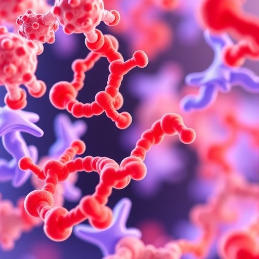
A groundbreaking advancement in the study of membrane proteins has been unveiled by researchers from Weill Cornell Medicine and Ruhr University Bochum, introducing an innovative fluorescence imaging-based technique that measures the activity rates of individual scramblase proteins. This novel methodology surpasses conventional ensemble approaches by providing an unprecedented, single-molecule resolution into the dynamics of scramblases—integral proteins responsible for lipid redistribution across cell membranes, which play pivotal roles in numerous biological processes.
Scramblases function by disrupting the asymmetrical distribution of lipids within the bilayer membrane, a phenomenon critical for cellular activities such as membrane assembly, protein glycosylation, programmed cell death, muscle development, and intracellular trafficking. Despite their biological significance, dissecting scramblase activity at the single-protein level has been an enduring challenge due to limitations inherent in bulk assays, which rely on averaging responses from populations of proteins and thus obscure the intrinsic heterogeneity of scramblase dynamics.
The innovative technique developed by the team leverages fluorescent tagging of scramblase proteins incorporated into synthetic lipid vesicles that mimic cell membranes. By immobilizing individual vesicles on glass slides and employing high-resolution fluorescence microscopy, the researchers could isolate vesicles harboring precisely one scramblase protein. This allowed for direct, quantitative measurements of lipid scrambling rates on a per-protein basis, revealing a vast spectrum of activities that were previously masked by ensemble averaging.
Focusing initially on the scramblase activity of VDAC1—a mitochondrial membrane channel recently discovered to possess scramblase function—the team found that VDAC1 operates as a dimeric complex with scrambling rates varying dramatically between individual protein pairs. These rates ranged from fewer than 100 to over 1,000 lipids translocated per second, highlighting a significant functional heterogeneity likely attributable to differing dimer conformations. These data provide molecular-level validation for computational models predicting conformer-dependent scramblase efficiency.
Expanding the application of their platform, the researchers examined opsin, a well-known G protein-coupled receptor in photoreceptor cells with an unexpected secondary role as a potent scramblase. Remarkably, individual opsin molecules exhibited lipid translocation rates exceeding 10,000 lipids per second, an order of magnitude greater than VDAC1 dimers. This discovery not only reinforces opsin’s functional versatility but also exemplifies the sensitivity and breadth of the new imaging method.
This fluorescence imaging-based platform offers profound flexibility for studying the influence of membrane composition, lipid environment, and pharmacological agents on scramblase function. By linking protein structure to activity through correlative high-resolution imaging, it becomes possible to elucidate the mechanistic underpinnings of scramblase regulation and dysfunction in human disease contexts.
Further ambitions for the technique include probing related lipid translocators such as flippases and floppases, proteins that also contribute to membrane lipid asymmetry but operate through distinct mechanisms. The capacity to measure individual protein activity within defined vesicular systems heralds a new era for membrane biology and drug discovery, enabling precise targeting of scramblase functions in pathological states.
The methodology’s advancement stands on the shoulders of pioneering ensemble assays originally developed by the Menon laboratory but catapults the field forward by circumventing their averaging limitations. This shift unlocks the ability to study scramblase functional heterogeneity, which may be critical for understanding the molecular basis of disorders linked to membrane lipid imbalances and for the design of scramblase-specific modulators.
The study exemplifies the power of interdisciplinary collaboration, intertwining biochemistry, biophysics, and advanced microscopy to elucidate membrane protein dynamics. It underscores the importance of technical innovation in revealing biological complexity at scales previously inaccessible, reinforcing the centrality of single-molecule approaches in modern biomedical research.
As scramblases emerge as promising therapeutic targets in a spectrum of diseases—from neurodegeneration to cancer—the availability of this cutting-edge single-protein assay platform could accelerate the identification of novel modulators, enhance mechanistic understanding, and ultimately contribute to precision medicine strategies that manipulate membrane lipid asymmetry for clinical benefit.
The findings of this seminal study, published in Nature Structural & Molecular Biology, reflect a significant leap forward in membrane protein research. By deciphering the kinetic variability and conformational dependencies of individual scramblase proteins, the work lays the groundwork for transformative research into the molecular machinery that governs cellular membrane architecture and function.
Subject of Research: Scramblase proteins; membrane lipid dynamics
Article Title: New single-protein fluorescence imaging technique reveals heterogeneous scramblase activity
News Publication Date: 15-Jun-2026
Web References:
- Menon Lab’s research on VDAC1 as a scramblase: https://www.nature.com/articles/s41467-023-43570-y
- Opsin’s dual function as a scramblase: https://www.sciencedirect.com/science/article/pii/S0960982210016994?via%3Dihub
References:
Nature Structural & Molecular Biology (Publication date: 15 June 2026)
Image Credits: Dr. Anant Menon
Keywords: Scramblase, lipid scrambling, VDAC1, opsin, single-protein analysis, fluorescence imaging, membrane proteins, biophysics, cell membrane dynamics, lipid transport, mitochondrial channels, molecular heterogeneity
