


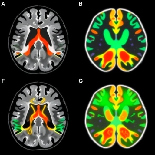
In a significant breakthrough that could redefine therapeutic strategies for Parkinson’s disease, researchers have unveiled how patient-specific brain connectivity analyses derived from ultra-high-field 7 Tesla MRI can enhance the precision and efficacy of deep brain stimulation (DBS). This pioneering approach leverages the unparalleled spatial and functional resolution provided by 7 Tesla magnetic resonance imaging to map the intricate neural circuits of individual patients, offering a tailored blueprint for neurosurgical targeting. The study, led by Wiggerts, Schuurman, de Bie, and colleagues and published in npj Parkinson’s Disease in 2026, exemplifies the convergence of cutting-edge imaging technology with precision neuromodulation, potentially transforming outcomes for the millions affected by Parkinson’s worldwide.
Deep brain stimulation has been instrumental in managing motor symptoms in Parkinson’s disease, particularly in advanced stages where pharmacological therapies falter. Despite DBS’s effectiveness, variability in patient outcomes persists, largely attributable to the challenges of identifying optimal stimulation targets within a highly interconnected and heterogenous brain network. Traditional targeting protocols primarily rely on standardized anatomical landmarks or indirect imaging modalities, which often fail to capture the nuanced variability of individual neural architecture. This new study confronts this obstacle head-on by harnessing the ultra-high spatial resolution of 7 Tesla MRI, capable of discerning microstructural connectivity patterns and functional linkages within the subcortical regions critical for Parkinsonian symptomatology.
By employing advanced diffusion tensor imaging (DTI) and resting-state functional MRI (rs-fMRI) sequences on the 7 Tesla platform, the researchers constructed comprehensive connectivity maps for each patient’s basal ganglia circuitry, specifically focusing on the subthalamic nucleus (STN) and globus pallidus internus (GPi), traditional DBS targets. Through sophisticated tractography and network connectivity analyses, they identified distinct pathways implicated in motor control, such as the hyperdirect pathway connecting the cortex to the STN, demonstrating individual variability in tract morphology, density, and functional coupling. These personalized connectomic profiles were instrumental in tailoring DBS electrode placement, shifting the practice from a “one-size-fits-all” paradigm to precision-guided neuromodulation.
The patient-specific targeting strategy yielded remarkable clinical outcomes, with treated individuals exhibiting significantly improved motor function as assessed by standardized scales like the Unified Parkinson’s Disease Rating Scale (UPDRS). This improvement was not merely incremental but surpassed results obtained through conventional targeting techniques, emphasizing the powerful synergy of high-field imaging and network neuroscience. Moreover, patients experienced a reduction in stimulation-induced adverse effects such as dysarthria and muscle contractions, which are often a consequence of inadvertent activation of neighboring fiber tracts. This highlights that connectivity-based targeting not only enhances efficacy but also optimizes safety by minimizing off-target stimulation.
A pivotal aspect of this technology-driven progress lies in the ability of 7 Tesla MRI to resolve microstructural features of the brain that earlier MRI field strengths could not reliably delineate. The ultra-high spatial resolution, often reaching submillimeter voxel sizes, enables visualization of the internal segmentation of subcortical nuclei and the intricate course of white matter fibers. The enhanced signal-to-noise ratio at 7T allows for more sensitive detection of subtle changes in tissue microstructure, improving the accuracy of diffusion metrics like fractional anisotropy and mean diffusivity. Collectively, these parameters feed into advanced computational models that predict optimal electrode trajectories and stimulation parameters tailored to individual patient anatomy and pathology.
Furthermore, the study underscores the indispensable role of network neuroscience in redefining neurological interventions. Parkinson’s disease is increasingly recognized as a network disorder, where dysfunction stems from maladaptive connectivity patterns across cortical and subcortical nodes rather than isolated regional abnormalities. Thus, the connectivity-informed DBS approach aligns with emerging concepts that target not only local sites but modulate entire functional networks to restore motor control and alleviate symptoms. This paradigm shift echoes broader trends in neuroscience where precision connectomics guides interventions from neurostimulation to rehabilitation.
The intricacies of this research involve integrating multimodal imaging data into surgical planning workflows. The team developed sophisticated software pipelines that combined anatomical, diffusion, and functional datasets, enabling neurosurgeons to visualize patient-specific brain connectivity directly overlaid on high-resolution structural images. These tools facilitated intuitive exploration of target sites in relation to critical pathways, supporting informed decision-making. The operators generated bespoke stimulation maps, fine-tuning electrode placement to maximize impact on motor network nodes while sparing critical circuits associated with cognitive and sensory function.
Equally remarkable is the translational potential of this advancement. As 7 Tesla MRI scanners become more widely available in clinical settings, the path to routine clinical adoption of connectomic DBS targeting appears increasingly plausible. The research paves the way for multicenter trials to validate the approach across diverse populations and imaging platforms. Additionally, the integration of machine learning algorithms to automate and refine connectivity analyses might further streamline preoperative planning, reducing time and resource burdens.
The implications extend beyond Parkinson’s disease, resonating with other movement disorders and neuropsychiatric conditions managed through neurostimulation. Patient-specific connectivity models could revolutionize treatment for essential tremor, dystonia, obsessive-compulsive disorder, and major depression, tailoring therapies to individual neural architecture. The convergence of ultra-high-field imaging, computational modeling, and neuromodulation heralds a new era of personalized brain medicine.
Moreover, the study highlights the importance of interdisciplinary collaboration, combining the expertise of neurologists, neurosurgeons, radiologists, physicists, and computational neuroscientists. This integrative approach was critical to overcoming technical challenges inherent in ultra-high-field imaging, such as susceptibility artifacts and RF inhomogeneities, which the team addressed through optimized pulse sequences and coil designs. The successful implementation of these technical innovations was central to realizing the clinical benefits reported.
Though promising, the approach does present challenges worthy of discussion. The high costs and limited accessibility of 7 Tesla MRI scanners remain barriers to widespread implementation, especially in under-resourced healthcare systems. Additionally, the increased scan time and complexity necessitate patient compliance and robust quality control protocols. Future research will need to address scalability and cost-effectiveness to ensure equitable access to these transformative techniques.
Interestingly, the study also validates earlier theoretical models positing that individualized neural circuitry underpins DBS outcomes. The direct evidence from patient-specific MRI data bridges gaps between preclinical animal models and human applications, reinforcing the biological plausibility of connectivity-driven interventions. This evidentiary chain strengthens confidence in pursuing personalized neuromodulation as a standard of care.
In conclusion, the groundbreaking work by Wiggerts et al. represents a milestone toward precision neuromodulation in Parkinson’s disease, leveraging the exquisite resolution of 7 Tesla MRI to decode individual brain connectivity and optimize deep brain stimulation. This study not only advances clinical neuroscience but also exemplifies how imaging technologies can transform patient care by tailoring therapies to unique neural fingerprints. As this methodology gains traction and expands, it promises to redefine therapeutic paradigms, improve quality of life, and inspire innovations across the neurological and psychiatric spectrum.
Cite Scienmag News
Cassandra Pierce. (May 28, 2026). 7T MRI Connectivity Enhances Parkinson’s DBS Targeting. Scienmag. https://scienmag.com/7t-mri-connectivity-enhances-parkinsons-dbs-targeting/
Cassandra Pierce. "7T MRI Connectivity Enhances Parkinson’s DBS Targeting." Scienmag, 28 May 2026, https://scienmag.com/7t-mri-connectivity-enhances-parkinsons-dbs-targeting/. Accessed 3 September 2026.
Cassandra Pierce. "7T MRI Connectivity Enhances Parkinson’s DBS Targeting." Scienmag. May 28, 2026. https://scienmag.com/7t-mri-connectivity-enhances-parkinsons-dbs-targeting/


