In a groundbreaking advancement in pulmonary medicine, researchers at Boston University Chobanian & Avedisian School of Medicine have revealed a novel framework that fundamentally redefines the understanding of pneumonia. This extensive study has unveiled seven distinct sub-phenotypes of pneumonia through detailed pulmonary histopathological examination—a method that looks beyond traditional diagnostic tools to scrutinize the microscopic cellular architecture and inflammation patterns within the lung tissue. This approach marks a pivotal shift away from the conventional reliance on symptomatic presentation, radiography, and microbial cultures, steering toward a personalized understanding of pneumonia’s diverse pathological manifestations.
Pneumonia, a formidable respiratory illness, consistently ranks among the leading causes of infectious mortality worldwide, particularly afflicting elderly populations in the United States. Historically, clinical strategies have generalized pneumonia treatment based on external signs, chest imaging results, and identification of causative pathogens. However, such methodologies neglect the intricate cellular and structural damage within the lungs that compound morbidity and residual dysfunction in survivors. This oversimplification has stymied targeted therapeutic advancements and masked the heterogeneity underlying pneumonia’s clinical variability.
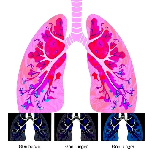
The Boston University study, published in the American Journal of Respiratory and Critical Care Medicine, harnessed state-of-the-art microscopy techniques alongside sophisticated machine learning algorithms to analyze autopsy lung samples from several hundred individuals who succumbed to pneumonia. By meticulously scoring twenty discrete types of histopathological abnormalities, spanning inflammation, alveolar damage, fibrosis, and immune infiltration, researchers identified seven reproducible clusters—each representing a unique pneumonia sub-phenotype. These clusters were characterized by distinct cellular infiltrates, damage patterns, and correlated variances in microbial agents.
This unprecedented classification delineates the multifaceted biological processes occurring within the pulmonary microenvironment during pneumonia. Some subgroups exhibited pronounced neutrophilic infiltration with widespread alveolar damage, indicative of aggressive inflammatory cascades. Others manifested predominant mononuclear immune cell accumulation coupled with fibrotic remodeling, suggestive of chronic or resolving injury phases. The diversity in lung tissue response elucidated via histopathology challenges the monolithic view of pneumonia and underscores the necessity for tailored clinical interventions addressing specific pathobiological pathways.
Linking these sub-phenotypes with the underlying microbiological milieu and immune signatures further validates their biological significance. Remarkably, similar histopathological patterns emerged in experimental mammalian models, corroborating the translational potential of these findings. This cross-species consistency provides a robust platform for mechanistic studies to unravel the etiological drivers and therapeutic vulnerabilities of each pneumonia subtype. It paves the way for innovative model systems that replicate human pneumonia heterogeneity with unprecedented fidelity.
Corresponding author Dr. Joseph P. Mizgerd, Professor of Pulmonary Medicine and director of the Pulmonary Center, emphasizes the novelty of subphenotyping pneumonia through pulmonary biology. He elucidates that pinpointing unique inflammatory and tissue damage signatures offers a strategic avenue to uncover biomarkers reflecting these sub-phenotypes in living patients—an essential step toward personalized diagnostics. The ultimate goal is to develop host-directed therapies calibrated to the specific biological perturbations shaping each pneumonia form.
The implications of this refined pneumonia taxonomy are profound. Current pneumonia treatment regimens, predominantly antibiotic-centric and symptomatic, do not adequately address the varied host immune responses and tissue damage patterns documented in this study. By stratifying patients based on underlying pathophysiology rather than solely on microbial identification or clinical symptoms, therapeutic precision may dramatically improve. This approach holds promise to mitigate long-term sequelae such as compromised pulmonary function, fibrosis, and accelerated respiratory decline frequently observed in older pneumonia survivors.
Moreover, this histopathologically driven framework offers critical insight into pneumonia’s contribution to unhealthy aging. The persistent lung damage and altered immune landscapes mapped by the researchers likely accelerate chronic respiratory diseases such as asthma and chronic obstructive pulmonary disease (COPD). Understanding these distinct trajectories will be instrumental in devising preventive strategies and rehabilitative interventions aimed at preserving lung health in aging populations disproportionately affected by pneumonia.
Future research trajectories, as outlined by the investigative team, involve validating peripheral biomarkers capable of non-invasively reporting these lung sub-phenotypes in clinical settings. Additionally, dissecting the molecular mechanisms orchestrating each histopathological variant will unveil novel therapeutic targets. Crucially, clinical trials will be necessary to assess whether host-targeted therapies yield differential efficacy across identified pneumonia subgroups, potentially revolutionizing treatment paradigms.
This seminal study not only redefines pneumonia classification but also exemplifies the power of integrating cutting-edge histopathology with machine learning analytics in respiratory medicine. The convergence of these technologies enables a granular understanding of lung disease complexity, fostering a new era of precision pulmonology. As respiratory infections remain a formidable global health challenge, such innovations offer hope for more effective, individualized interventions to reduce pneumonia’s substantial mortality and morbidity burden.
The research was supported by multiple grants from the National Institutes of Health and collaborations involving neurological and aging-related disease centers, emphasizing the interdisciplinary interest in pneumonia’s systemic impacts. The findings serve as a clarion call for the medical community to reassess current diagnostic and therapeutic algorithms, encouraging the adoption of biologically informed subphenotyping approaches that embrace pneumonia’s intricate pathobiology.
In sum, the Boston University researchers have illuminated a nuanced landscape of pneumonia pathology, challenging conventional wisdom and charting a course toward personalized pulmonary care. By recognizing that pneumonia comprises multiple distinct disease processes with unique cellular signatures and immune landscapes, this work lays the foundation for future breakthroughs in diagnosis, treatment, and ultimately, prevention of one of the world’s most persistent infectious threats.
Subject of Research: Cells
Article Title: Sub-phenotypes of pneumonia defined by pulmonary histopathological features
News Publication Date: 28-Apr-2026
Web References: 10.1093/ajrccm/aamag182
Keywords: Pneumonia, pulmonary histopathology, sub-phenotypes, lung inflammation, alveolar damage, machine learning, host-directed therapy, respiratory disease, immune profiles, personalized medicine, inflammation, tissue remodeling
