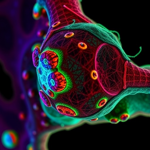
In the rapidly evolving field of super-resolution fluorescence microscopy, a pioneering breakthrough has been made by researchers from Harbin Institute of Technology and Peking University, addressing one of the most stubborn challenges in cellular imaging: background artifacts that confound quantitative analysis. The newly developed Adaptive Self-inspired Noise2Noise (Adaptive-SN2N) framework revolutionizes how noisy images are denoised, enabling unprecedented accuracy in visualizing and segmenting subcellular structures like mitochondria and the endoplasmic reticulum (ER) with remarkable clarity and reliability.
Fluorescence microscopy, essential for visualizing cellular dynamics, often faces the dilemma of balancing sufficient illumination with the risk of photobleaching and phototoxicity. To mitigate these risks during prolonged live-cell imaging, light exposure is reduced, resulting in low photon counts that degrade image quality via reduced signal-to-noise ratio (SNR). Conventional denoising approaches, including self-supervised deep learning models such as Noise2Noise and Noise2Void, have alleviated the need for clean reference images and demonstrated impressive photon efficiency. However, they are prone to generating artificial structures in signal-absent areas, misleading biological interpretations and subsequent computational analyses.
The core problem arises from standard preprocessing techniques—particularly the commonly employed “patch-wise normalization.” This approach intensifies subtle, random noise in background regions by amplifying local statistical discrepancies, which neural networks then erroneously interpret as genuine biological features. For images containing sparse biological signals or predominantly dark backgrounds, these artifacts manifest as false positives in downstream processes like organelle segmentation or synaptic pattern recognition, undermining the scientific validity of the findings.
To tackle this, the research team embarked on an interdisciplinary exploration combining expertise in optical super-resolution imaging, algorithmic deep learning, and cell biology. Their approach hinged on a rigorous mathematical analysis that revealed how patch-wise normalization inflates noise variance in low dynamic range patches by an alarming factor proportional to the inverse square of the patch’s local intensity range (ΔP). This normalization effect stretches subtle background noise across the normalized intensity interval [0,1], creating fabricated structures. High-intensity sparse patches simultaneously undergo compression of background pixels into negligible intensity ranges, causing significant shifts in data distributions that challenge robust denoising.
The Adaptive-SN2N framework introduces a paradigm shift through a risk-aware adaptive normalization strategy. By quantitatively evaluating each image patch’s statistical risk profile—factoring in mean intensity, standard deviation, and skewness—the algorithm dynamically selects the optimal normalization format. For high-risk patches where weak backgrounds or sparse bright signals prevail, image-wise global normalization replaces traditional patch-wise methods to prevent over-amplification of noise and distorted data distributions. Conversely, for low-risk patches containing dense structural information, localized patch-wise normalization is retained to maximize contextual contrast, preserving fine detail fidelity.
Complementing this, Adaptive-SN2N employs an innovative self-inspired learning regimen leveraging a single noisy image. Through diagonal spatial resampling combined with Fourier domain interpolation, the framework synthesizes twin image pairs sharing identical biological content but independent noise realizations. This clever dual-image generation negates the need for clean ground-truth references, facilitating a self-constrained training process that hones denoising performance while protecting against artifact creation. This strategy exemplifies the advancing frontier of unsupervised and self-supervised machine learning tailored for biological imaging.
Furthermore, the team addressed boundary discontinuities and stitching artifacts inherent in patch-based inference by implementing a Gaussian-weighted overlap across sliding windows. Utilizing a 50% overlap rate combined with smooth 2D Gaussian weighting functions, predicted image segments are seamlessly integrated, eliminating abrupt transitions and ensuring spatial coherence. Exhaustive ablation studies confirmed that this approach significantly enhances denoising quality across multiple datasets and diverse metrics, underscoring its robust generalizability.
Experimental validation on both structured illumination microscopy (SIM) and spinning-disk SIM platforms demonstrated Adaptive-SN2N’s superior capability in handling complex live-cell dual-color imaging. The technique prevented extensive false-segmentation in mitochondrial background regions during Otsu thresholding, a common automated segmentation pitfall. It also dramatically improved the morphological continuity of the ER skeleton from an extensively fragmented baseline to a cohesive, topologically faithful network. This fidelity enabled precise tracking of dynamic mitochondrial fission and fusion events, crucial for understanding cellular physiology and pathology.
Crucially, the Adaptive-SN2N framework delivers these performance gains without compromising photon efficiency, maintaining improvements of one to two orders of magnitude compared to conventional methods, while fundamentally suppressing background artifacts at their source. This represents a theoretical and practical milestone in computational microscopy, reconciling the competing demands of noise suppression, structural authenticity, and phototoxicity mitigation.
Looking forward, the principles of risk-aware adaptive processing pioneered here promise transformative impact across a broad spectrum of computational microscopy tasks beyond denoising. Anticipated applications include enhanced image segmentation, precise organelle colocalization analysis, and the rigorous assessment of subcellular interactions. Particularly in neuroscience, artifact-free, high-SNR imaging will facilitate high-density synaptic tracking, while in oncology, it will enable detection of minute metastatic protrusions with unprecedented reliability.
By eradicating deceptive artifacts that previously confounded image interpretation, Adaptive-SN2N opens new avenues for quantitative life science research built on a foundation of data authenticity and reproducibility. As imaging resolutions and analytical complexity continue to escalate, this landmark framework stands poised to underpin a new generation of discoveries at the interface of biology, optics, and machine intelligence.
The ingenuity of Adaptive-SN2N lies in its seamless fusion of theoretical insight, methodological innovation, and biological relevance, setting a new gold standard for artifact-suppressed super-resolution imaging. As self-supervised learning frameworks gain traction, this adaptive, risk-aware approach highlights the critical importance of preprocessing strategies in safeguarding the scientific integrity of computational microscopy outputs, ensuring that the images we see indeed represent the truth within living cells.
Subject of Research: Cells
Article Title: Artifact-suppressed and adaptive self-inspired learning denoising for super-resolution fluorescence microscopy
News Publication Date: 31-Mar-2026
Web References: http://dx.doi.org/10.3724/PXLIFE.2025-0010
Image Credits: Harbin Institute of Technology / Jingyang Zhu
Keywords: super-resolution microscopy, fluorescence microscopy, deep learning denoising, adaptive normalization, noise reduction, mitochondrial imaging, endoplasmic reticulum, self-supervised learning, computational microscopy, photobleaching, image segmentation, artifact suppression
