In a groundbreaking study addressing one of the most insidious challenges in geriatric medicine, researchers have unveiled critical insights into the risk factors associated with intracranial injuries in older adults who suffer minor head trauma. This investigation dives deep into the importance of cutaneous lesion localization—a nuanced and often overlooked element that could revolutionize diagnostic precision and therapeutic interventions for this vulnerable demographic.
Traumatic brain injuries (TBIs), even when classified as minor, pose a significantly heightened threat to the elderly population, given their increased susceptibility due to cerebral atrophy, vessel fragility, and comorbidities. The study by Selki, Demir, and Şengüldür navigates the complex terrain of identifying which external manifestations—specifically cutaneous lesions—serve as reliable prognostic markers for underlying intracranial pathology.
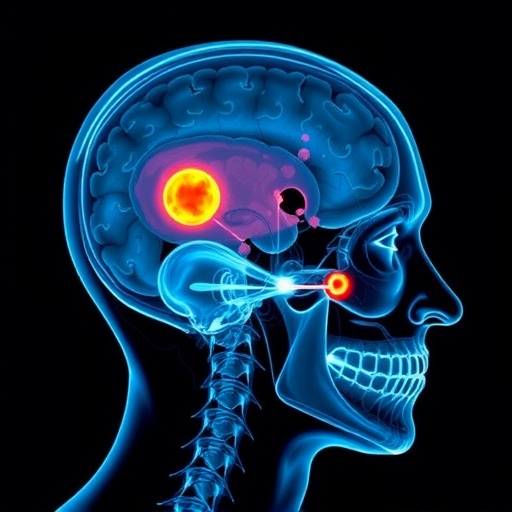
One of the pivotal revelations emerging from this research is the spatial correlation between the site of cutaneous trauma and the risk probability of intracranial hemorrhages or contusions. Unlike in younger cohorts, where diffuse impact might be the norm, older adults demonstrate a distinctive pattern whereby lesions localized in certain cranial regions serve as red flags, signaling a more ominous risk profile beneath the skin’s surface.
The clinical implications of these findings cannot be overstated. Historically, minor head trauma in seniors often results in conservative management with limited imaging, primarily due to cost constraints or the clinical presentation’s subtleness. However, the identification of lesion localization as a predictive tool necessitates a paradigm shift, advocating for targeted diagnostic imaging protocols such as CT scans or MRIs, expressly when lesions appear in high-risk zones.
Furthermore, the study systematically dissects the influence of coexisting medical conditions, including anticoagulant therapy—a commonality in the elderly—that exacerbates vulnerability to intracranial bleeding. The interplay between external lesion location and such systemic factors paints a more comprehensive risk stratification model, enabling physicians to calibrate urgency and interventions meticulously.
Methodologically, Selki and colleagues employed a robust cohort study design, rigorously analyzing demographic data, lesion phenotypes, anatomical distribution, and subsequent imaging outcomes. The granularity of their dataset enabled statistically significant correlations, surpassing previous research attempts that typically homogenized lesion sites or conflated injury severities.
One of the most striking elements of the research is its challenge to long-held clinical heuristics which treated minor head trauma in older adults with a broad-brush approach. This study’s data advocate for tailored assessments that prioritize lesion mapping to refine clinical suspicion for intracranial injury, potentially reducing both missed diagnoses and unnecessary radiological exposure.
Moreover, the pathophysiological underpinnings explored in the discussion lend credence to why certain cranial regions are predisposed to more severe underlying damage. Variations in skull thickness, regional cerebral blood flow dynamics, and structural vulnerabilities collectively contribute to these findings, informing not only diagnostic pathways but potential preemptive protective strategies in eldercare.
The research also highlights a compelling need for enhanced education among emergency care providers regarding cutaneous lesion significance. Broad-based training could improve early identification of high-risk presentations, expediting specialist referrals, and optimizing patient outcomes in a cohort often marginalized in acute trauma protocols.
In terms of public health implications, the insights provided by this study underscore the urgency of revising existing clinical guidelines to incorporate lesion localization assessments as a core component of head trauma evaluation in older adults. Implementing these recommendations could lead to substantial improvements in morbidity and mortality statistics associated with traumatic brain injuries in this age group.
Importantly, the study also tackles limitations inherent in current diagnostic frameworks, such as reliance on Glasgow Coma Scale scores potentially masking subtle but critical injuries in elderly patients. It advocates adopting novel adjunctive measures, with lesion location serving as an accessible yet powerful parameter to augment traditional assessments.
Interdisciplinary collaboration emerges as a crucial pathway forward, with neurologists, radiologists, emergency physicians, and geriatric specialists working synergistically to translate these findings into clinical practice. Tailoring treatment algorithms to this nuanced risk factor could also stimulate innovation in protective gear design, rehabilitation techniques, and preventive health strategies.
Future research directions, inspired by this study, might encompass longitudinal analyses tracking outcomes relative to initial lesion locations, integrating technological advancements such as machine learning to predict injury trajectories. Enhancing predictive analytics within this framework promises to elevate precision medicine in geriatric trauma care.
Beyond the immediate clinical sphere, ethical considerations surface, especially regarding healthcare resource allocation and balancing risks of imaging overuse against life-saving diagnostics. This research enriches the dialogue with empirical data supporting more judicious, evidence-based decision-making in complex eldercare scenarios.
In the broader context of population aging globally, where the incidence of falls and minor head traumas is rising, this study acts as a clarion call for healthcare systems to anticipate and adapt services accordingly. Strategic investments in training, diagnostic technologies, and public awareness campaigns centered on lesion localization insights could significantly mitigate the burden of traumatic brain injuries.
Ultimately, this pioneering research redefines the landscape of minor head trauma in older adults by emphasizing a seemingly peripheral yet profoundly impactful factor: the precise location of cutaneous lesions. The potential to transform clinical vigilance and patient outcomes by integrating this knowledge marks a notable advance in geriatric trauma management.
As the medical community continues to grapple with the complexity of brain injuries in aging populations, the insights delivered by Selki, Demir, and Şengüldür provide an invaluable compass. They highlight that, within the subtle contours of the skin’s surface, lies critical information offering a clearer window into the brain’s hidden vulnerabilities.
The intersection of meticulous clinical observation and rigorous scientific inquiry, as demonstrated here, embodies the future of personalized, efficacious medical care—especially for those in their most vulnerable years. In this frame, cutaneous lesion localization transcends its modest facade to become a cornerstone in safeguarding older adults from the silent crisis of intracranial injuries.
Subject of Research: Risk factors for intracranial injuries in older adults following minor head trauma, with a focus on cutaneous lesion localization.
Article Title: Risk factors for intracranial injuries in older adults with minor head trauma: how important is cutaneous lesion localization?
Article References:
Selki, K., Demir, M.C. & Şengüldür, E. Risk factors for intracranial injuries in older adults with minor head trauma: how important is cutaneous lesion localization? BMC Geriatr 26, 410 (2026). https://doi.org/10.1186/s12877-025-06853-1
Image Credits: AI Generated
