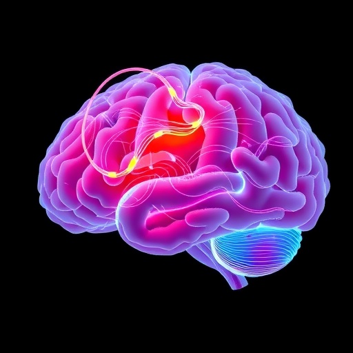
In an unprecedented breakthrough study published in The Lancet Digital Health, researchers from the USC Mark and Mary Stevens Neuroimaging and Informatics Institute have uncovered remarkable insights into how the brain’s architecture radically reorganizes itself following stroke-induced impairments. This international collaborative effort reveals that, paradoxically, while the hemisphere directly damaged by the stroke undergoes accelerated aging, the corresponding regions on the opposite side exhibit signs of a younger, more resilient brain structure. This counterintuitive finding illuminates the brain’s extraordinary capacity for adaptation and plasticity, reshaping long-held assumptions about stroke recovery.
This study was propelled by an internationally coordinated project under the Enhancing NeuroImaging Genetics through Meta-Analysis (ENIGMA) Stroke Recovery Working Group. Researchers meticulously examined brain MRI data from over 500 stroke survivors drawn from 34 research centers spanning eight countries. Employing cutting-edge deep learning algorithms trained on extensive neuroimaging datasets, they refined methods to estimate the “brain age” of discrete cerebral regions within each hemisphere. This regional brain age estimation enabled the team to discern nuanced differences in structural integrity that conventional imaging techniques failed to detect.
Associate Professor Hosung Kim, a neuroscientist and co-senior author, highlighted the compelling nature of these findings, noting that larger strokes not only exacerbate biological aging of the injured hemisphere but also elicit an unexpected rejuvenation effect in the contralesional brain regions. This rejuvenation likely reflects the brain’s effort to compensate for lost motor functions by enhancing undamaged networks. These dynamics underscore the complex interplay of neural degeneration and compensatory plasticity during chronic stages of stroke recovery.
Central to the methodology was the application of a graph convolutional network, an advanced artificial intelligence model adept at parsing the intricate connectivity patterns of the brain. This approach enabled the extraction of localized brain age estimates from MRI scans, measured against chronological age to yield the brain-predicted age difference (brain-PAD). Brain-PAD served as a sensitive biomarker indicating the varying degrees of neural health and degeneration across stroke-affected and unaffected brain areas.
Notably, the contralesional frontoparietal network—an essential hub for motor planning, attention, and coordination—showed significantly younger brain age profiles in stroke survivors exhibiting severe and persistent motor impairments. This suggests that rather than achieving full functional recovery, the unaffected hemisphere undergoes structural reorganization aimed at mitigating deficits. This finding challenges the traditional binary view of stroke recovery and positions neuroplasticity as a multifaceted process with potential markers detectable through advanced neuroimaging.
This study underscores the power of aggregating neuroimaging data across global cohorts to overcome the limitations of smaller, isolated studies that may miss subtle, yet clinically meaningful, brain adaptations. By integrating data from diverse populations and using artificial intelligence to decode complex brain patterns, researchers are opening new frontiers in personalized neurology. These insights hold promise for tailoring individualized rehabilitation strategies that leverage a patient’s unique neural plasticity profile.
Arthur W. Toga, director of the Stevens Institute and a co-author, emphasized the transformative potential of this research. He explained that the revealed heterogeneity in brain aging post-stroke could serve as a roadmap to predict recovery trajectories, thereby informing targeted interventions. Future clinical protocols may incorporate brain-PAD metrics as a diagnostic adjunct, evolving rehabilitation from a generalized approach to one finely tuned to each patient’s neurobiological state.
Plans are underway to expand the scope of investigation with longitudinal studies tracking patients from acute stroke phases through chronic recovery stages. Monitoring the temporal dynamics of brain age changes will shed light on the evolution of compensatory neuroplasticity and could identify critical windows where interventions might be most efficacious. This time-resolved analysis aims to transform stroke recovery management by enabling truly adaptive therapies.
Additionally, the study highlights the enormous value of interdisciplinary collaboration, blending neurology, radiology, computational neuroscience, and artificial intelligence. This confluence of expertise facilitated the development of novel analytical frameworks capable of penetrating the complexities of stroke pathology and brain adaptation. As a result, the research represents a paradigm shift in how clinicians conceptualize and measure brain health after trauma.
In the broader context of cerebrovascular disorders, these discoveries contribute to a growing body of evidence that brain aging is not uniform, but rather regionally heterogeneous, especially in pathological conditions. The revelation that neuroplastic processes manifest as region-specific rejuvenation challenges existing clinical dogma and opens avenues for research into other neurological diseases where compensatory mechanisms might be similarly exploited.
This landmark study was supported by funding from the National Institutes of Health and engaged collaborators from leading global institutions including the University of British Columbia, Monash University, Emory University, and the University of Oslo. The extensive data harmonization and methodological rigor demonstrate the feasibility and power of multinational neuroimaging consortia like ENIGMA to address complex neurological questions.
Ultimately, this research reframes the narrative around stroke recovery, positioning the brain not just as a victim of injury but as an active agent of structural reformation and resilience. By quantifying region-specific brain aging and identifying contralesional neuroplasticity, the study paves the way for innovative diagnostic tools and personalized rehabilitation, transforming patient care and offering hope to millions affected by stroke worldwide.
Subject of Research: People
Article Title: Associations between contralesional neuroplasticity and motor impairment through deep learning-derived MRI regional brain age in chronic stroke (ENIGMA): a multicohort, retrospective, observational study
News Publication Date: 22-Jan-2026
Web References: https://enigma.ini.usc.edu/, https://www.thelancet.com/journals/landig/article/PIIS2589-7500(25)00124-4/fulltext
References: Deep learning prediction of MRI-based regional brain age reveals contralesional neuroplasticity associated with severe motor impairment in chronic stroke: A worldwide ENIGMA study, The Lancet Digital Health, DOI: 10.1016/j.landig.2025.100942
Image Credits: Stevens INI
Keywords: Brain, Brain injuries, Brain structure, Neuroimaging, Cerebrovascular disorders, Neuroplasticity, Artificial intelligence, Ischemia
