A revolutionary leap in biomedical imaging and anatomical research has arrived with the launch of an extraordinary open-access 3D portal known as the Human Organ Atlas (HOA). This emerging platform grants users unprecedented access to exquisitely detailed three-dimensional renderings of human organs, permitting interactive exploration from the macro scale of an entire organ to near-cellular resolution at the micron level. Harnessing a cutting-edge imaging method, Hierarchical Phase-Contrast Tomography (HiP-CT), the Atlas combines the capabilities of the European Synchrotron Radiation Facility’s (ESRF) newly commissioned Extremely Brilliant Source—remarkably brighter than conventional CT imaging systems—with novel computational techniques to produce datasets that verge on the microscopic. This innovation not only transforms anatomical visualization but carves new paths in medical research, education, and artificial intelligence-driven diagnostics.
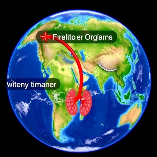
The Human Organ Atlas opens a fresh window into the intricate architecture of human organs such as the brain, heart, lungs, kidneys, and liver. By leveraging the HiP-CT methodology, intact ex vivo organs are scanned without destructive preparation, preserving their complex three-dimensional microstructures. HiP-CT bridges a longstanding gap between the conventional realms of clinical radiology and microscopic histology—two fields traditionally disparate in their spatial and resolution scales. Where traditional CT captures millimetric anatomical features and histology focuses on cellular details from thin tissue slices, HiP-CT spans this continuum. This synchrotron-based imaging produces volumetric data with resolutions down to less than one micron, enabling scientists and clinicians to “fly through” organ landscapes, exploring them layer by layer with unparalleled clarity.
Developed through an international collaboration spearheaded by University College London (UCL) in partnership with the ESRF and other esteemed institutes across Europe and the United States, the Human Organ Atlas Hub represents a monumental coordinated effort in open, interdisciplinary science. Over five years in the making, the project integrates expertise from biomedical imaging, mechanical engineering, clinical research, and data science, unified in a vision to democratize access to the highest-quality organ datasets ever generated. The initiative emphasizes FAIR data principles—making datasets Findable, Accessible, Interoperable, and Reusable—ensuring worldwide availability without the need for specialised hardware or software.
The advent of the Extremely Brilliant Source at ESRF has been pivotal. This next-generation synchrotron light source provides photon flux intensities up to 100 billion times stronger than hospital-grade CT scanners, enormously enhancing image contrast and resolution. HiP-CT capitalizes on this by capturing hierarchical imaging datasets: starting from whole-organ scans encompassing billions of voxels down to microscale volumes exposing cellular and subcellular structures. The data sizes are colossal, with individual organ datasets often spanning hundreds of gigabytes, sometimes exceeding a terabyte, with the most massive brain dataset nearing a staggering 14 terabytes.
This groundbreaking platform is not confined to niche academic circles; it is designed for broad accessibility. Interactive web-based viewers allow users to navigate these vast datasets seamlessly through standard browsers. Alongside visualization, comprehensive tools and tutorials facilitate analysis and interpretation, equipping researchers, educators, and students alike to engage deeply with human anatomy and pathophysiology. The portal perpetually evolves, with new datasets and tools regularly released to expand the atlas’s scope and utility.
Scientifically, the Human Organ Atlas is already propelling medical understanding forward. Initially conceived amid the COVID-19 pandemic, the project’s imaging prowess has shed light on previously undetectable vascular injuries within infected lungs, altering clinical perceptions of viral pathogenesis. Furthermore, applications in cardiac research have revisited and refined interpretations of heart disease manifestations. Insights into complex gynecological disorders, such as adenomyosis, have likewise benefitted from the atlas’s ability to visualize uterine vasculature in three dimensions, revealing microanatomical features invisible to earlier technologies.
Clinician-scientists like Judith Huirne from the Amsterdam UMC emphasize the immense value of virtual 3D histological data for unraveling disease mechanisms and addressing deep-seated gender disparities in medical research. These volumetric reconstructions empower pathologists and medical professionals with a holistic view of organs and tissues, potentially revolutionizing diagnostics and therapeutic strategies. By enabling more precise structural-to-functional correlations, the Human Organ Atlas fosters an integrative understanding of human health and disease.
From an educational perspective, the HOA is transformative. Traditional anatomy instruction, often reliant on static images and physical specimens, can now be supplemented with immersive digital exploration. Students and educators gain the ability to virtually dissect and analyze organs interactively, fostering spatial intelligence and a more intuitive grasp of physiological structures and their interrelations. This dynamic mode of learning enhances retention and engagement, modernizing curricula and public outreach alike.
The datasets generated through HiP-CT hold exceptional promise in the rapidly evolving field of artificial intelligence. High-resolution, volumetric, and well-curated medical imaging datasets are critical for training machine learning models designed for automated segmentation, anomaly detection, and super-resolution imaging. The Human Organ Atlas offers a rare, standardized resource for developing and validating AI tools that could augment diagnostic accuracy and personalized medicine. By bridging gaps between imaging scales, the atlas also paves the way for multiscale modeling of organ function and disease progression.
Though already vast, the Human Organ Atlas is only the beginning of a longer journey toward comprehensive digital biobanks. The consortium envisions expanding the repository to encompass a broader array of organs, diverse donor samples, and richer metadata. Future technical developments aspire to scale HiP-CT imaging to complete human bodies with resolutions surpassing current capabilities by an order of magnitude, potentially reshaping our conceptual and practical approaches to anatomy and medicine. This bold ambition underscores a commitment to open science and global collaboration.
Behind the scenes, the Human Organ Atlas Hub exemplifies extraordinary interdisciplinary teamwork. Engineers, imaging experts, clinician-scientists, data managers, and infrastructure specialists collaborate intensively to optimize imaging protocols, data processing pipelines, and user interfaces. ESRF scientists like Paul Tafforeau play a central role in evolving and applying the HiP-CT technology, while UCL’s team, led by professors Peter Lee and Claire Walsh, anchors much of the biological and engineering research integration. The collective expertise and vision driving this project embody the transformative potential of “team science” in the biomedical frontier.
As the Human Organ Atlas grows in scale and impact, it promises to redefine paradigms across biomedical research, clinical practice, education, and technology development. By providing unparalleled access to the microanatomy of intact human organs, this platform invites scientists and the public alike to explore the mysteries of human biology in revolutionary ways. In an era hungry for breakthroughs in understanding disease and optimizing health, the alliance of cutting-edge imaging technology with open data democratization heralds a new epoch for medicine and science.
Subject of Research: Not applicable
Article Title: The Human Organ Atlas
News Publication Date: 11-Mar-2026
Web References: http://dx.doi.org/10.1126/sciadv.adz2240
References:
1. Walsh CL et al., Imaging intact human organs with local resolution of cellular structures using hierarchical phase-contrast tomography, Nat. Methods 18, 1532–1541 (2021).
2. Brunet J et al., Multidimensional Analysis of the Adult Human Heart in Health and Disease Using Hierarchical Phase-Contrast Tomography, Radiology 312(1), e232731 (2024).
3. HOAHub Uterine Consortium, Revealing the unseen: 3D synchrotron X-Ray imaging of uterine vasculature in adenomyosis, Angiogenesis 29(1), 3 (2026).
Image Credits: UCL data led ESRF
Keywords: Human Organ Atlas, Hierarchical Phase-Contrast Tomography, HiP-CT, Synchrotron Imaging, Extremely Brilliant Source, Biomedical Imaging, 3D Organ Visualization, Medical AI, Anatomy Education, COVID-19 Pathology, Cardiac Imaging, Gynecological Disorders
