In an exciting breakthrough that promises to transform the landscape of live-cell imaging, researchers Martin Küppers and W.E. Moerner have unveiled an innovative microscopy technique known as Interferometric Image Scanning Microscopy (I-ISM). Published February 27, 2026, in the journal Light: Science & Applications, this cutting-edge method achieves an unprecedented lateral resolution of 120 nanometers inside living cells without relying on fluorescent labels. This advancement addresses long-standing challenges in cellular biology and microscopy, offering scientists a powerful new tool for exploring the intimate architecture of life at the nanoscale.
Traditional optical microscopy techniques have always wrestled with inherent physical limits governed by the diffraction of light, typically capping lateral resolution at around 200-250 nanometers. Fluorescence microscopy, especially super-resolution variants like STED and PALM/STORM, has previously breached this threshold by tagging cellular structures with fluorescent probes. However, these methods carry significant drawbacks, including photobleaching, phototoxicity, and the potential to perturb natural cellular behavior due to labeling. The advent of label-free imaging at nanoscale resolution opens vast new vistas for observing biological processes in their pristine, unmodified states.
Interferometric Image Scanning Microscopy builds upon the foundational concept of image scanning microscopy (ISM), which merges the principles of confocal microscopy with a detector array to enhance resolution. By integrating interferometric detection schemes, Küppers and Moerner have ingeniously exploited the phase information of light waves scattered or emitted by cellular structures. This phase-sensitive detection markedly boosts signal-to-noise ratios and spatial resolution, while simultaneously preserving the viability of live cells under observation.
The core innovation lies in the delicate orchestration of interferometric signal acquisition with point-scanning illumination. By scanning a focused laser beam across the sample and collecting emitted or backscattered light via an interferometer, the technique extracts high-fidelity spatial data. This dual-detection approach captures both amplitude and phase data of the light field, enabling computational reconstruction of cellular ultrastructure with lateral precision reaching 120 nm—roughly twice as sharp as conventional confocal microscopy, but vastly gentler than super-resolution fluorescence techniques.
Most strikingly, I-ISM achieves this remarkable resolution without the dependency on fluorescent dyes, sidestepping the intrinsic challenges of labeling live specimens. Label-free imaging is especially critical in delicate cellular contexts, such as stem cell differentiation or dynamic protein complex formation, where exogenous tags may interfere with native biological behaviors. In these scenarios, the ability to visualize nanoscale cellular features in vivo, unencumbered by artifacts, opens revolutionary opportunities for real-time biological discovery.
Another compelling aspect of this technique revolves around its adaptability to thick biological tissues. Fluorescence microscopy’s limitations in-depth penetration and phototoxic effects are well documented, often necessitating invasive sample preparation or fixation. With I-ISM, researchers can maintain native physiological conditions while probing deep within three-dimensional living tissues, extending nanoscale imaging capabilities substantially deeper than previously achievable with label-dependent methods.
The technical implementation of I-ISM represents a tour de force in optical instrumentation. The researchers employ a highly stable interferometric setup combined with precision scanning optics and sensitive detection arrays. Such integration demands rigorous optical alignment, phase stabilization, and sophisticated computational algorithms for phase retrieval and image reconstruction. The system’s exquisite sensitivity captures subtle optical path length differences reflected by cellular nanostructures, enabling the dissection of organelle morphology, cytoskeletal frameworks, and membrane dynamics with newfound clarity.
Beyond technical specifications, the implications for biological research are profound. For instance, visualizing the dynamic arrangement of chromatin within the nucleus, tracking intracellular transport vesicles, or monitoring mitochondrial morphology changes, all become feasible with unparalleled clarity and temporal resolution. This can catalyze discoveries in cellular physiology, disease pathology, and drug response mechanisms by offering a real-time window into nanoscale transactions that dictate cellular fate.
The potential clinical applications of label-free I-ISM are equally compelling. Non-invasive, high-resolution imaging inside living human tissues could revolutionize diagnostic procedures, enabling early detection of pathological alterations at the molecular level without biopsies or labeling agents. Additionally, this method could guide precision surgery or targeted therapeutic delivery by providing surgeons and clinicians with critical structural insights during interventions.
Küppers and Moerner’s pioneering work continues the trajectory of advancing microscopy into realms once thought inaccessible due to physical constraints. Their fusion of interferometric principles with image scanning microscopy exemplifies how classical optical physics can be revitalized to meet contemporary biomedical challenges. This synergy of physics, engineering, and biology underscores the evolving nature of interdisciplinary science driving innovation.
Moreover, this advancement dovetails nicely with ongoing developments in computational imaging and artificial intelligence. The rich datasets produced by I-ISM stand to benefit from AI-driven image analysis tools that can extract meaningful biological insights from complex interferometric patterns. This confluence of hardware and software innovations paves the way for automated, high-throughput nanoscale imaging pipelines.
While the current demonstration focuses on lateral resolution improvements, future adaptations of I-ISM might enhance axial resolution and even enable volumetric imaging at sub-diffraction limits. Combining interferometric phase detection with light-sheet microscopy or adaptive optics could further alleviate scattering and aberrations, broadening applicability across diverse biological specimens.
In addition to biological sciences, the principles underlying I-ISM may find resonance in materials science, nanotechnology, and semiconductor diagnostics where nanoscale surface characterization is paramount. The label-free and non-destructive nature of the technique positions it as a versatile tool beyond life sciences, facilitating precise imaging of nanoscale phenomena in a variety of technical domains.
In conclusion, the advent of Interferometric Image Scanning Microscopy marks a monumental step forward in the pursuit of label-free, ultrahigh-resolution imaging inside living cells. By circumventing the traditional constraints of fluorescence labeling and diffraction limits, Küppers and Moerner have equipped researchers with a transformative microscope capable of unveiling the nuanced nano-architecture of life with unprecedented fidelity. As this technology matures and proliferates, it promises to reshape our understanding of cellular dynamics, accelerate biomedical discoveries, and inspire novel technological innovations across disciplines.
Subject of Research: Development of a label-free super-resolution microscopy method for live-cell imaging.
Article Title: Interferometric Image Scanning Microscopy for label-free imaging at 120 nm lateral resolution inside live cells.
Article References:
Küppers, M., Moerner, W.E. Interferometric Image Scanning Microscopy for label-free imaging at 120 nm lateral resolution inside live cells. Light Sci Appl 15, 129 (2026). https://doi.org/10.1038/s41377-026-02210-y
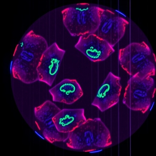
Image Credits: AI Generated
DOI: 27 February 2026
