


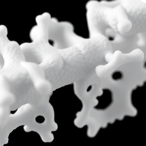
A laboratory model developed by scientists at the Indian Institute of Science is offering researchers a more realistic way to study why some triple-negative breast cancers respond poorly to chemotherapy. The experimental system uses a light-printed material that changes shape after fabrication, transforming from a flat sheet into a narrow, tubular structure when immersed in liquid. By recreating both the geometry of a breast duct and the gentle movement of fluid through living tissue, the model reveals how physical conditions surrounding cancer cells can influence their activity, organization and response to treatment.
Triple-negative breast cancer is considered one of the most aggressive forms of breast cancer because its cells lack three commonly targeted receptors: estrogen receptors, progesterone receptors and human epidermal growth factor receptor 2, or HER2. Because these molecular targets are absent, treatments that work for many other breast cancers are not effective against this subtype. Chemotherapy remains an important treatment option, but tumors can develop resistance or respond unevenly. Understanding the physical and biological conditions that shape this behavior is therefore a major challenge for cancer researchers.
Traditional laboratory studies often grow cancer cells on flat plastic surfaces, where they form a thin layer exposed to a relatively uniform environment. That arrangement is convenient for experiments, but it differs substantially from the three-dimensional architecture of a tumor inside the body. Breast cancer cells exist within tissue that has curvature, mechanical support, nearby extracellular matrix and movement of fluids. These factors can affect how cells receive nutrients, remove waste, communicate with one another and encounter therapeutic drugs. The new platform was designed to bring several of those influences into a single laboratory model.
The researchers created the structure through a form of four-dimensional bioprinting. In this context, the fourth dimension refers to a programmed change in shape over time rather than simply the addition of another spatial direction. Light is used to print a material into a defined initial geometry. Once the printed construct is placed in liquid, internal stresses and the material’s programmed response cause the flat sheet to fold into a tube. The resulting duct-like structure resembles the curved, enclosed spaces in which some breast cancers originate, while also providing a controlled environment in which researchers can position and observe living cells.
The team introduced triple-negative breast cancer cells into the printed tubes and examined them under two different conditions. In the first, the cultures remained stationary, providing a conventional static environment. In the second, the tubes were gently moved on a rocker to imitate the low-level fluid motion that can occur in biological tissues. The rocking did not reproduce every feature of blood flow or the complex circulation of fluids in a tumor. Instead, it created a carefully controlled mechanical stimulus, allowing the researchers to compare cancer-cell behavior in the presence or absence of movement while keeping the surrounding experimental conditions as similar as possible.
The cancer cells remained highly viable in both settings, indicating that the printed material and tubular architecture could support their survival. Yet viability alone did not capture the most important difference between the cultures. Cells exposed to dynamic conditions showed greater metabolic activity, suggesting that fluid movement altered their energy use or physiological state. They also changed shape and organization within the tube. Such changes matter because a cancer cell’s geometry and arrangement can influence how it interacts with neighboring cells, attaches to its surroundings and responds to signals from the tissue environment.
The most striking result emerged when the researchers exposed the cultures to doxorubicin, a widely used chemotherapy drug. Cells grown under dynamic conditions showed greater survival after treatment than cells maintained in static culture. This finding suggests that fluid movement and tissue architecture may contribute to a more treatment-resistant state, even when the cancer cells themselves are genetically similar. The observation does not mean that rocking a laboratory culture directly reproduces drug resistance in a patient. Rather, it demonstrates that mechanical and structural conditions can change the way cancer cells respond to an established therapy, potentially affecting the results of drug-screening experiments.
The study also highlights why three-dimensional models are increasingly important in cancer research. A flat culture can help scientists measure cell growth, toxicity and molecular responses, but it may miss interactions created by curvature, confinement and spatial organization. The 4D-printed tubes allow these variables to be studied together with dynamic stimulation. Researchers can potentially modify the dimensions of the structure, alter the surrounding material or introduce other cell types to examine how the tumor microenvironment affects disease progression. The platform could also support experiments that compare drug concentrations, treatment schedules and combinations of therapies under more tissue-like conditions.
The model remains an experimental research tool rather than a clinical treatment or a substitute for testing in patients. It contains a simplified population of cancer cells and does not fully reproduce the immune system, blood vessels, hormonal signals or the diverse cell populations found in a human tumor. Its findings will need to be confirmed through additional laboratory studies and, ultimately, more clinically relevant models. Even so, the work provides a valuable warning against treating static, flat cultures as complete representations of cancer biology. By showing that movement and shape can influence both cell behavior and chemotherapy survival, the researchers have created a platform that may help explain why promising treatments sometimes perform differently in living tissue than they do in conventional laboratory dishes.
Web References: https://doi.org/10.1016/j.engreg.2026.07.002
References: Engineered Regeneration, DOI: 10.1016/j.engreg.2026.07.002
Cite Scienmag News
Nathaniel Bowman. (August 15, 2026). 4D-Printed Breast Cancer Model Mimics Ducts, Revealing Treatment Resistance. Scienmag. https://scienmag.com/4d-printed-breast-cancer-model-mimics-ducts-revealing-treatment-resistance/
Nathaniel Bowman. "4D-Printed Breast Cancer Model Mimics Ducts, Revealing Treatment Resistance." Scienmag, 15 August 2026, https://scienmag.com/4d-printed-breast-cancer-model-mimics-ducts-revealing-treatment-resistance/. Accessed 4 September 2026.
Nathaniel Bowman. "4D-Printed Breast Cancer Model Mimics Ducts, Revealing Treatment Resistance." Scienmag. August 15, 2026. https://scienmag.com/4d-printed-breast-cancer-model-mimics-ducts-revealing-treatment-resistance/


