


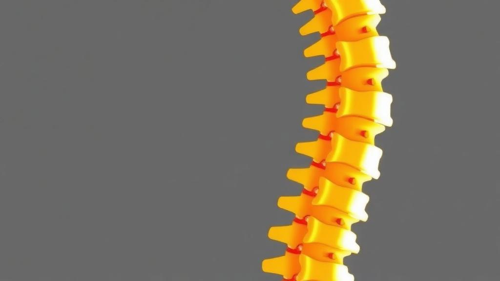
When a landmine or improvised explosive device detonates beneath a military vehicle, the blast wave hurls the floor upward and drives a violent vertical shock through the seat, the pelvis, and ultimately the lumbar spine. The result is a distinctive and devastating injury pattern: compression and burst fractures of the lower back vertebrae, which in severe cases can crush the spinal cord and leave survivors with permanent impairment, spinal deformity, and chronic pain. Yet despite decades of research into battlefield trauma, the biomechanics community has lacked a fundamental piece of information — how fracture tolerance varies from one lumbar vertebra to the next. A new combined experimental and computational study published in the Annals of Biomedical Engineering has now delivered the first vertebral level-specific injury prediction curves for the lumbar spine under high-rate vertical loading, and the findings could reshape how protective systems for soldiers are designed.
The research, led by Kwong Ming Tse of Swinburne University of Technology and the University of Melbourne, together with Dale Robinson, Melanie Franklyn of Australia’s Defence Science and Technology Group, and Peter Vee Sin Lee of the University of Melbourne, tackled a problem that has long frustrated injury biomechanics researchers. Most existing spinal injury criteria were derived from tests on entire spinal columns or large spinal sections, which provide useful system-level tolerance estimates but reveal little about which individual vertebra is most likely to fail. Earlier work had hinted at the importance of this question. Studies of vertical accelerations simulating aircraft ejection showed that as acceleration increases, injuries migrate from the thoracolumbar junction down toward the lower lumbar spine. Sled experiments simulating underbelly blast found that longer-duration seat pulses of 55 milliseconds reduced spinal responses by 68 to 78 percent compared with shorter 10-millisecond pulses, producing fewer pelvic and spinal injuries. But none of these approaches could isolate the vulnerability of each lumbar level.
To fill this gap, the team took an unusual two-pronged approach. They obtained five cadaveric lumbar spine specimens, each free of severe abnormalities as confirmed by radiography and visual inspection, and used dual-energy X-ray absorptiometry to verify that all specimens had areal bone mineral density values within the normal range reported for young adult males aged 17 to 25 years, despite the donors being under 62 years of age. From each spine they isolated a three-vertebra segment — a central vertebra flanked by its neighbours and two intervertebral discs — with the outer vertebrae partially embedded in dental plaster potting fixtures and the middle vertebra deliberately left exposed. Each segment was then compressed at approximately one metre per second on a mechanical testing machine, a rate chosen to bridge conventional quasi-static testing and the more severe blast-level conditions, where reported vehicle floor velocities in live-fire blast events range from 2.2 to 12.8 metres per second. The researchers note that the velocity actually experienced by the lumbar spine is lower than the floor velocity because the seat, pelvis, and surrounding soft tissues attenuate the load.
Alongside the physical tests, the team built specimen-specific finite element models of each three-vertebra segment from high-resolution computed tomography scans with a pixel size of just 0.098 millimetres. The models were anatomically meticulous: vertebral bodies were discretised with roughly 145,000 tetrahedral elements each, cortical shells were divided into seven regions with distinct properties, intervertebral discs comprised ten annulus layers surrounding a nucleus pulposus modelled with hyperelastic material laws, and all seven major spinal ligaments were represented as membrane elements. Crucially, the CT data were used to assign bone material properties element by element, using a calibration phantom to convert Hounsfield units to bone density and a power-law regression to derive Young’s modulus for the trabecular bone. When the models were run in an explicit finite element solver replicating the compression experiments, they reproduced the measured force–displacement responses with coefficients of determination between 0.93 and 0.98, and predicted fracture loads within 10 to 34 percent of the experimental values. The models also captured the observed failure patterns, including coronal split fractures of the vertebral body, impaction fractures of the superior endplate, and isolated trabecular fractures — morphologies consistent with those reported clinically in underbelly blast casualties.
With the models validated, the researchers unleashed them on a series of parametric simulations that would be impossible to perform experimentally. They varied spinal posture by adding 10 degrees of flexion or extension, mimicking the range of sagittal intervertebral rotation during maximal seated trunk movement, and added 10 degrees of lateral bending comparable to voluntary side-bending. They scaled vertebral height and cross-sectional area by plus or minus 10 percent, reflecting anthropometric variation across a military population. And they swept areal bone mineral density across a physiologically relevant range of 0.8 to 1.4 grams per square centimetre, corresponding to young healthy soldiers. The results revealed a striking level-dependence that no whole-column experiment could have exposed.
Posture emerged as a powerful, level-specific modulator of fracture risk. At L1, L2, and L4, flexion generally reduced the likelihood of injury, shifting the fracture probability curves rightward, while extension increased the risk. At L3 the trend reversed, and at L5 both flexion and extension reduced fracture tolerance — a unique behaviour the authors attribute to the biomechanical complexity of the lowest lumbar vertebra, including its adjacency to the sacrum and its altered facet joint geometry. Lateral bending increased fracture susceptibility at the lower lumbar levels, particularly L5, where the decreased fracture load coincided with a marked increase in vertebral body loading. The stiffness analysis added another layer of nuance: at the L4–L5–S1 segment, flexion cut the compressive load at 5 millimetres of displacement from 6,545 newtons to 2,499 newtons, a 62 percent reduction. Importantly, changes in axial stiffness did not consistently translate into changes in fracture load, underscoring that stiffness and strength are distinct metrics that both matter when assessing injury tolerance.
Geometry and bone quality told a more consistent story. Increasing vertebral height reduced axial stiffness and fracture load at all levels except L5, where the opposite trend appeared — a finding that contrasts with earlier ex vivo work reporting a positive correlation between vertebral height and strength, and one the authors suggest may reflect biological factors such as bone mineral density and trabecular connectivity not captured in the models. Larger cross-sectional area, by contrast, consistently enhanced mechanical resistance at every level. Areal bone mineral density proved to be a strong and reliable positive predictor of compressive fracture tolerance across the entire lumbar spine, with higher density producing systematic increases in both stiffness and failure load. Yet the analysis produced a surprise: a measure called axial rigidity, derived from CT-based geometry and material properties, actually outperformed bone mineral density as a predictor of injury risk, yielding injury curves with narrow confidence intervals that converged toward a single vertebra-independent relationship.
The practical implications reach well beyond the laboratory. Fracture tolerance generally increased from L1 to L4, tracking the increasing size and strength of the vertebrae, but L5 consistently bucked the trend, remaining highly vulnerable despite its larger dimensions. This persistent weakness identifies the lower lumbar spine as a critical target for injury mitigation — a finding that could guide the design of vehicle seats, armour, and blast-attenuating flooring. The injury prediction curves, generated using parametric survival analysis with a Weibull distribution, also align closely with injury corridors previously developed for whole lumbar spine segments, lending credibility to the modelling approach. The authors caution that their fracture tolerance values should be read as vertebral-level injury metrics rather than direct operational thresholds, since the experiments isolated axial compression and did not replicate the full combined-loading environment of a real blast, and the experimental component involved one specimen per lumbar level owing to the scarcity of suitable cadaveric material.
Even with those limitations, the study marks a genuine milestone. By integrating spinal posture, vertebral geometry, and bone quality into validated, specimen-specific finite element models, the researchers have created tools for injury prediction that are both anatomically specific and clinically relevant. The framework extends naturally beyond the battlefield: the same biomechanics governs spinal loading in aviation ejections, helicopter crashes, and occupational impact scenarios. And the finding that axial rigidity may be a more robust predictor of vertebral fragility than the bone density measurements routinely used in clinics hints at a future where fracture risk assessment becomes more precise — not just for soldiers facing blast threats, but for anyone whose spine must withstand the sudden, merciless forces of a high-rate impact.
Subject of Research: Level-specific biomechanical tolerance of lumbar vertebrae to high-rate vertical compression relevant to underbelly blast injury
Article Title: Level-Specific Lumbar Spine Injury Tolerance Under High-Rate Vertical Loading Relevant to Underbelly Blast Environments: A Combined Cadaveric Experimental and Finite Element Study of Three-Vertebra Spinal Segments
Article References: Tse, K. M., Robinson, D., Franklyn, M., & Lee, P. V. S. (2026). Level-Specific Lumbar Spine Injury Tolerance Under High-Rate Vertical Loading Relevant to Underbelly Blast Environments: A Combined Cadaveric Experimental and Finite Element Study of Three-Vertebra Spinal Segments. Annals of Biomedical Engineering. https://doi.org/10.1007/s10439-026-04347-8
Image Credits: AI Generated
DOI: 10.1007/s10439-026-04347-8
Keywords: lumbar spine, underbelly blast, finite element model, cadaveric testing, fracture tolerance, bone mineral density, spinal posture, injury prediction curves, military biomechanics, burst fractures, vertebral geometry, axial rigidity
Cite Scienmag News
Ophelia Keating. (October 3, 2026). Which Lumbar Vertebra Breaks First in a Blast? New Study Maps Spine Fracture Risk Level by Level. Scienmag. https://scienmag.com/which-lumbar-vertebra-breaks-first-in-a-blast-new-study-maps-spine-fracture-risk-level-by-level/
Ophelia Keating. "Which Lumbar Vertebra Breaks First in a Blast? New Study Maps Spine Fracture Risk Level by Level." Scienmag, 3 October 2026, https://scienmag.com/which-lumbar-vertebra-breaks-first-in-a-blast-new-study-maps-spine-fracture-risk-level-by-level/. Accessed 4 October 2026.
Ophelia Keating. "Which Lumbar Vertebra Breaks First in a Blast? New Study Maps Spine Fracture Risk Level by Level." Scienmag. October 3, 2026. https://scienmag.com/which-lumbar-vertebra-breaks-first-in-a-blast-new-study-maps-spine-fracture-risk-level-by-level/


