


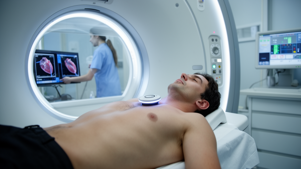
When doctors want to measure how stiff the heart muscle is, they can use a technique called cardiac magnetic resonance elastography, or MRE. It works by vibrating the chest from the outside and tracking how the resulting shear waves travel through the heart. Because stiffer tissue carries waves differently than softer tissue, the technique can quantify myocardial stiffness without a biopsy. But the method has a weak link that has received surprisingly little attention: where, exactly, the vibrating device is placed on the chest. A new study from the Department of Radiology at the First Affiliated Hospital of the First Affiliated Hospital of Anhui Medical University, published in Frontiers in Frontiers, set out to answer that question directly.
Cardiac MRE fills a diagnostic gap that conventional imaging leaves open. Echocardiography, cardiac CT, and standard cardiac MRI can suggest that the heart muscle has changed — through wall thickness measurements, strain patterns, or T1 mapping values — but none of these tools measures stiffness itself. Instead, they offer indirect surrogates that can be confounded by factors unrelated to the true mechanical properties of the myocardium. Elastography, by contrast, interrogates the tissue directly: an external driver generates low-frequency mechanical vibrations, a passive device on the chest wall transmits those vibrations into the thorax, and phase-contrast MRI captures the shear waves as they propagate through the beating heart. Specialized reconstruction algorithms then convert the wave field into spatially resolved stiffness maps, all without contrast agents or invasive pressure measurements.
The clinical motivation for such a direct measurement is substantial. Increased myocardial stiffness is now recognized as an early and prognostically important feature of a range of cardiac diseases, including myocardial fibrosis, hypertrophic cardiomyopathy, cardiac amyloidosis, restrictive cardiomyopathy, and heart failure with preserved ejection fraction. Detecting these changes before they become irreversible could shape treatment decisions and improve long-term outcomes. Cardiac MRE has already demonstrated the ability to detect elevated myocardial stiffness in cardiac amyloidosis, hypertrophic cardiomyopathy, aortic stenosis, and pulmonary hypertension, and its measurements have been shown to correlate with invasive hemodynamic indices. This track record has led many investigators to regard cardiac MRE as a quantitative imaging biomarker with genuine translational potential.
Yet the technique faces a fundamental physical challenge that distinguishes it from its more established cousin, liver MRE. The liver sits close to the abdominal wall and offers a relatively uniform acoustic pathway for mechanical waves. The heart, by contrast, lies behind the ribs and is partially surrounded by air-filled lung tissue, which attenuates and scatters mechanical vibrations before they can reach the myocardium. The heart also moves rapidly throughout the cardiac cycle and has a complex three-dimensional geometry, both of which complicate the delivery of consistent wave energy to the left ventricular wall. In the single-frequency protocol used in this study, mechanical excitation at one vibration frequency was delivered through a single passive driver, which meant that the driver’s position on the chest wall determined where shear waves entered the thorax and how efficiently they reached the left ventricular myocardium.
Despite this obvious dependence on driver placement, most published cardiac MRE studies have positioned the driver at the apical surface projection — the region to the left of the sternum and just above the xiphoid process — based largely on practical convention rather than on direct comparative evidence. The left chest wall, located at the same axial level as the apical surface projection but along the mid- to anterior-axillary line, represents a clinically feasible alternative when anterior placement is difficult. Such difficulties are not rare in practice: prior thoracic surgery, implanted cardiac devices, breast tissue, obesity, and chest wall deformities can all make anterior placement problematic or impossible. The left chest wall is close to the heart, accessible in the supine position, and compatible with the same fixation band used for conventional placement. The right chest wall, by comparison, sits farther from the heart, and posterior placement is poorly suited to routine supine cardiac MRI workflows. Alternative left lateral placement is therefore a practical and recurring clinical consideration rather than a hypothetical concern.
To compare the two positions objectively, the researchers needed a quantitative measure of wave quality rather than relying on subjective image inspection alone. They turned to the octahedral shear strain signal-to-noise ratio, a metric commonly used in elastography to characterize shear-wave quality. This measure reflects how clearly the propagating waves stand out against background noise in the strain field, and it has been shown to be associated with the reliability of stiffness estimation. When OSS-SNR is low, the inversion algorithms that convert wave data into stiffness values become less robust, and measurement variability tends to increase. A recent normative study of 125 healthy adults had already identified acquisition-related factors — including driver coupling and slice location — as important sources of variability in cardiac MRE, and other investigators had explicitly flagged actuator placement as a key but understudied determinant of wave quality. What was missing was a direct, within-subject comparison of driver positions.
The new study provided exactly that. Twenty-three healthy volunteers with a mean age of 43 years underwent two consecutive cardiac MRE acquisitions at 3.0 Tesla during a single session. The passive driver was placed first at the apical surface projection and then moved to the left chest wall at the same axial level, with the scan repeated after re-localization. Participants were asked to avoid strenuous exercise and stimulant-containing food or drink on the day of scanning, and each rested supine for at least thirty minutes before the examination to limit transient blood pressure swings. Left ventricular myocardial stiffness and OSS-SNR were measured at both positions, and differences were analyzed using paired statistical tests appropriate for within-subject comparisons.
The results were striking. Myocardial stiffness measured at the apical surface projection averaged 9.0 kPa, compared with 14.7 kPa at the left chest wall — a mean difference of 5.8 kPa, which corresponds to a very large effect size. This finding carries an important implication: the two driver positions do not simply differ in image quality, they yield systematically different stiffness values in the same healthy hearts. Any study or clinical program that switches driver positions could introduce a shift in measured stiffness that is large relative to the differences expected between health and disease. Standardizing driver placement is therefore not merely a matter of convenience but a prerequisite for comparing results across studies, institutions, and time points.
The wave quality data help explain why the two positions diverge. OSS-SNR was significantly higher at the apical surface projection than at the left chest wall, again with a substantial effect size. Higher wave quality at the anterior position plausibly reflects a shorter and less obstructed path for mechanical vibrations traveling from the chest surface to the heart, with less attenuation by lung tissue along the way. Better wave delivery in turn supports more reliable stiffness inversion, which is consistent with the reproducibility findings.
Reproducibility was assessed by having observers repeat the stiffness measurements and by comparing measurements between independent observers, using intraclass correlation coefficients and Bland–Altman analysis. At the apical surface projection, stiffness measurements demonstrated excellent agreement both within and between observers, with intraclass correlation coefficients of 0.962 and 0.963 respectively. At the left chest wall, intra-observer agreement remained excellent at 0.914, but inter-observer agreement dropped to 0.839, a level generally classified as good rather than excellent. The pattern suggests that when wave quality degrades, different observers analyzing the same images begin to diverge more in their stiffness estimates, likely because noisier wave fields leave more room for interpretive judgment during region-of-interest placement and quality control.
For clinicians and researchers, the practical takeaway is that the apical surface projection should be considered the preferred driver position for cardiac MRE, offering both higher wave quality and superior reproducibility. The left chest wall remains a viable fallback when anterior placement is impractical, but its use should be documented explicitly, and stiffness values obtained at the two positions should not be interpreted interchangeably. The magnitude of the observed difference — nearly 6 kPa in healthy volunteers — underscores how consequential this seemingly technical choice can be.
Several aspects of the study design merit attention when considering how the findings generalize. The cohort consisted exclusively of healthy volunteers, so the behavior of wave propagation in diseased hearts — with altered geometry, remodeled walls, or pericardial constraints — remains to be established. The sample size of twenty-three, while adequate for a within-subject comparison with large effect sizes, is modest, and the participants spanned a wide age range. Future work will need to determine whether the superiority of the apical position holds in patient populations, across different vibration frequencies, and with the multi-driver or multi-frequency configurations that some cardiac MRE protocols employ. Nevertheless, by directly comparing two clinically realistic driver positions within the same subjects, the study converts a matter of convention into a matter of evidence, and it provides a concrete, actionable standard for anyone seeking to add cardiac MRE to clinical or research practice.
Subject of Research: Cardiac Imaging
Article Title: Evaluating the impact of passive driver position on image consistency and repeatability in 3.0T cardiac magnetic resonance elastography
Article References: Evaluating the impact of passive driver position on image consistency and repeatability in 3.0T cardiac magnetic resonance elastography. (n.d.). https://doi.org/10.3389/fradi.2026.1912855
Image Credits: AI Generated
DOI: 10.3389/fradi.2026.1912855
Keywords: cardiac MRE, myocardial stiffness, reproducibility, apical surface, lateral surface, imaging biomarkers, Evaluating, impact, passive, driver, position, image
Cite Scienmag News
Ophelia Keating. (October 15, 2026). Where You Place the Vibrator Changes What the Heart MRI Reads: New Study Pins Down the Best Spot. Scienmag. https://scienmag.com/where-you-place-the-vibrator-changes-what-the-heart-mri-reads-new-study-pins-down-the-best-spot/
Ophelia Keating. "Where You Place the Vibrator Changes What the Heart MRI Reads: New Study Pins Down the Best Spot." Scienmag, 15 October 2026, https://scienmag.com/where-you-place-the-vibrator-changes-what-the-heart-mri-reads-new-study-pins-down-the-best-spot/. Accessed 15 October 2026.
Ophelia Keating. "Where You Place the Vibrator Changes What the Heart MRI Reads: New Study Pins Down the Best Spot." Scienmag. October 15, 2026. https://scienmag.com/where-you-place-the-vibrator-changes-what-the-heart-mri-reads-new-study-pins-down-the-best-spot/


