


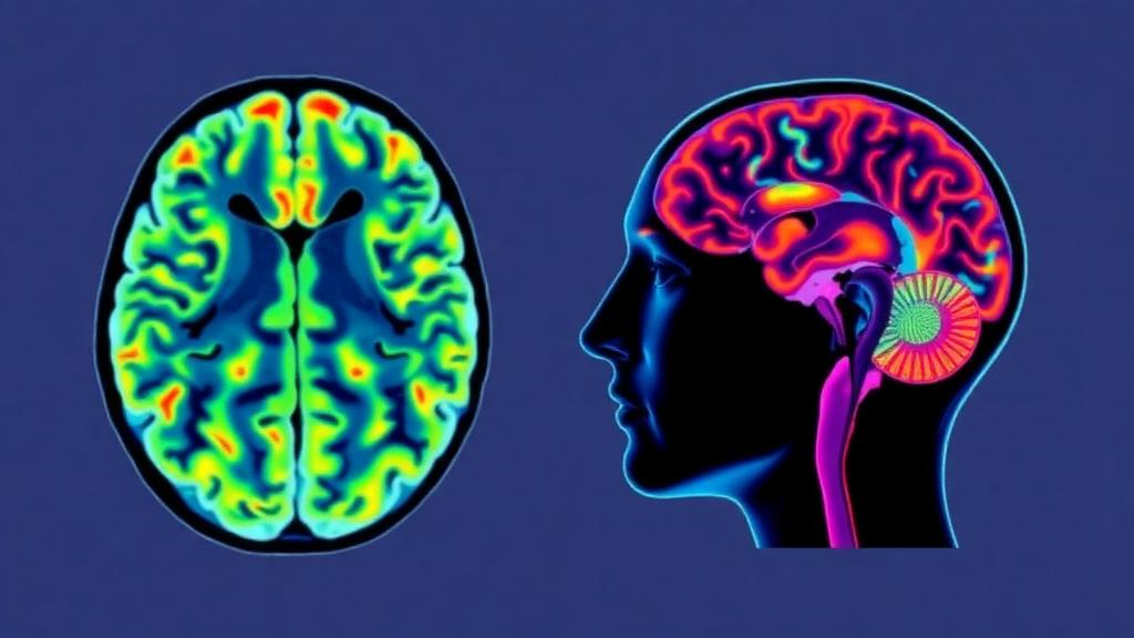
What happens to blood flow inside the brain when the heart is suddenly pushed to beat faster and harder? A team of researchers in Beijing set out to answer that question with a technique that requires no contrast agents, no radiation, and no invasive catheters: arterial spin labeling, or ASL, a magnetic resonance imaging method that uses magnetically tagged water in the blood as its own tracer. In an exploratory study published in BMC Medical Imaging, Tingting Zhang of Beihang University and colleagues measured cerebral blood flow estimates in healthy young volunteers both at rest and during dobutamine stress, a pharmacological challenge that mimics the hyperdynamic circulation of intense exercise. The findings offer a cautious but intriguing glimpse into how the brain’s perfusion landscape may shift when cardiovascular drive surges, while also highlighting the considerable technical and practical challenges of imaging the brain under physiological stress.
The study enrolled 52 volunteers aged 23 to 33 years, of whom four were excluded because of contraindications to MRI or dobutamine administration. From the remaining 48 eligible participants, only 21 ultimately completed all required acquisitions. The reason for the substantial drop-out is telling: 27 volunteers could not finish the second of the study’s two arterial spin labeling acquisitions because of discomfort during the prolonged dobutamine stress examination. Dobutamine, a synthetic catecholamine-like drug, is a mainstay of cardiac stress testing, but lying still inside a noisy MRI scanner while a drug pushes your heart rate and blood pressure upward is far from pleasant. The attrition underscores a practical reality of stress physiology research: the human body under pharmacological strain does not always cooperate with the demands of high-resolution imaging.
The technical heart of the study lies in a parameter known as the post-labeling delay, or PLD. In pseudo-continuous arterial spin labeling, or pCASL, blood water in the neck arteries is magnetically labeled, and after a waiting period, images are acquired. The labeled water has by then exchanged with tissue water, and the resulting signal difference can be converted into an estimate of cerebral blood flow in milliliters per 100 grams of tissue per minute. The choice of PLD is critical. If the delay is too short, the labeled blood may not yet have reached the imaging slices, particularly in regions with long arterial transit times, leading to underestimated flow. If it is too long, the label decays away. The researchers used two sequential single-PLD three-dimensional pCASL acquisitions at delays of 1500 milliseconds and 2500 milliseconds, always in the same fixed order, at 3.0 Tesla during both rest and dobutamine stress.
Dobutamine did exactly what it was expected to do to the body. The infusion significantly increased heart rate, systolic blood pressure, diastolic blood pressure, mean arterial pressure, and respiratory rate, while oxygen saturation remained stable. This constellation of changes defines the hyperdynamic state the investigators sought to create, one in which cardiac output rises and the entire circulatory system operates under heightened drive. The question was whether the brain, which is famously protected by cerebral autoregulation, a mechanism that keeps perfusion remarkably constant across swings in systemic blood pressure, would show measurable changes in its blood flow estimates at all, and if so, where and under which imaging timing.
The answer, at the shorter delay, was essentially no. At PLD equal to 1500 milliseconds, none of the twelve region-of-interest comparisons between rest and stress reached even the nominal unadjusted threshold of P less than 0.05. In other words, when the labeled blood was sampled relatively soon after labeling, the ASL-derived cerebral blood flow estimates looked statistically indistinguishable between the resting and hyperdynamic states across the whole brain, tissue masks, lobes, and vascular territories. This is consistent with the classical view of cerebral autoregulation holding the line, but it may also reflect the physics of the measurement itself, since a shorter delay is more vulnerable to errors when arterial transit times change, as they plausibly do when cardiac output and blood pressure surge.
At the longer delay of 2500 milliseconds, a different picture emerged. Nominal unadjusted increases in ASL-derived cerebral blood flow estimates were observed in three regions: the temporal lobe, the territory supplied by the posterior cerebral artery, and the vertebrobasilar territory, which encompasses the posterior circulation fed by the vertebral and basilar arteries. Only the temporal lobe showed a nominal condition-by-PLD interaction, meaning its response differed between the two delays, while the posterior cerebral artery and vertebrobasilar territories did not reach that level of interaction. The authors were careful to frame these as exploratory observations, noting that the fixed sequential acquisition order and the absence of statistical correction for multiple comparisons mean these regional patterns cannot be taken as established evidence of tissue-specific or delay-dependent responses.
Beyond the regional maps, the team explored whether the change in whole-brain cerebral blood flow, expressed as delta CBF, correlated with the physiological shifts induced by dobutamine. At the 1500-millisecond delay, delta CBF showed a nominal correlation with the change in heart rate. At 2500 milliseconds, nominal correlations appeared with both heart-rate change and diastolic-pressure change. These relationships hint at a coupling between the magnitude of the cardiovascular challenge and the perfusion signal captured by ASL, but the authors emphasize that such exploratory correlations require confirmation with properly powered, counterbalanced designs before any physiological interpretation can be considered firm.
The methodological caveats deserve attention because they shape what the study can and cannot claim. The tissue-mask cerebral blood flow values were derived without individual structural segmentation or partial-volume correction and were treated as purely descriptive. The two PLD acquisitions were always performed in the same order, so any systematic drift in physiology, drug effect, or scanner stability over time is confounded with the delay itself. The authors explicitly state that the findings do not establish PLD-dependent or tissue-specific responses, nor the superiority of a longer post-labeling delay. What the study does establish is a feasible, if demanding, protocol for acquiring ASL data during pharmacological stress, along with a set of hypothesis-generating observations about posterior circulation and temporal lobe perfusion under hyperdynamic conditions.
Why does this matter beyond the laboratory? Arterial spin labeling is increasingly used to study cerebrovascular reactivity, the brain’s capacity to adjust its blood supply, which is relevant to conditions ranging from hypertension and diabetes to dementia and, intriguingly, spaceflight. The study was supported in part by the Space Medical Experiment Project of China’s Manned Space Program, reflecting a growing interest in how the cardiovascular and cerebrovascular systems behave under altered physiological loads. A validated non-contrast method for probing brain perfusion during cardiovascular stress could eventually complement established tools such as blood-oxygenation-level-dependent cerebrovascular reactivity mapping, transcranial Doppler ultrasound, and dynamic susceptibility contrast MRI, each of which has its own limitations in stress settings.
The authors call for further studies using counterbalanced multi-PLD imaging, direct assessment of arterial transit time, and synchronized physiological monitoring to clarify how much of the observed change reflects true perfusion shifts versus sampling timing artifacts. Until then, the study stands as a careful, honest first step: a demonstration that ASL can be attempted during dobutamine stress in humans, that the brain’s perfusion estimates largely hold steady under autoregulation at shorter sampling delays, and that longer delays may reveal regional changes in the posterior circulation that merit rigorous follow-up. For a field where the brain and the heart are usually studied separately, work that watches them interact in real time, inside the magnet, points toward a more integrated physiology of the circulation under stress.
Subject of Research: ASL-derived cerebral blood flow measurement during dobutamine stress in healthy adults
Article Title: ASL-derived cerebral blood flow estimates during dobutamine stress in healthy subjects: an exploratory study using two sequential single-PLD acquisitions
Article References: Zhang, T., Cheng, X., Cai, L., Zhang, S., Niu, H., Yang, Z., Wang, Z., Ren, P., & Liu, Y. (2026). ASL-derived cerebral blood flow estimates during dobutamine stress in healthy subjects: an exploratory study using two sequential single-PLD acquisitions. BMC Medical Imaging. https://doi.org/10.1186/s12880-026-02898-9
Image Credits: AI Generated
DOI: 10.1186/s12880-026-02898-9
Keywords: arterial spin labeling, cerebral blood flow, dobutamine stress, post-labeling delay, pCASL, cerebral autoregulation, hyperdynamic circulation, posterior circulation, MRI, arterial transit time, cerebrovascular reactivity, BMC Medical Imaging
Cite Scienmag News
Cassandra Pierce. (October 13, 2026). When the Heart Races, the Brain’s Blood Flow Map Shifts: MRI Study Tracks Cerebral Circulation Under Drug-Induced Stress. Scienmag. https://scienmag.com/when-the-heart-races-the-brains-blood-flow-map-shifts-mri-study-tracks-cerebral-circulation-under-drug-induced-stress/
Cassandra Pierce. "When the Heart Races, the Brain’s Blood Flow Map Shifts: MRI Study Tracks Cerebral Circulation Under Drug-Induced Stress." Scienmag, 13 October 2026, https://scienmag.com/when-the-heart-races-the-brains-blood-flow-map-shifts-mri-study-tracks-cerebral-circulation-under-drug-induced-stress/. Accessed 13 October 2026.
Cassandra Pierce. "When the Heart Races, the Brain’s Blood Flow Map Shifts: MRI Study Tracks Cerebral Circulation Under Drug-Induced Stress." Scienmag. October 13, 2026. https://scienmag.com/when-the-heart-races-the-brains-blood-flow-map-shifts-mri-study-tracks-cerebral-circulation-under-drug-induced-stress/


