


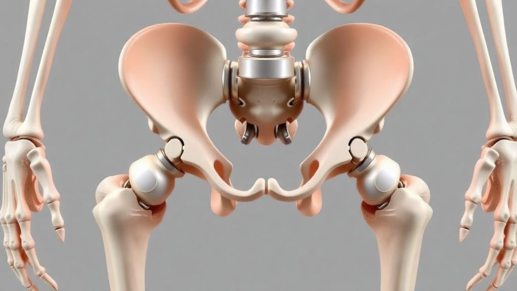
Total hip replacement is one of the most common and most successful operations in modern medicine, and its popularity is only set to grow as populations age and healthcare systems expand. Among the surgical techniques available, the direct anterior approach, or DAA, has earned a devoted following because it lets surgeons work through the front of the hip with the patient lying on their back, without cutting through muscle. Patients typically get back on their feet faster, and the risk of the new joint dislocating backwards is lower. But the technique has a demanding secret: it requires the surgeon to pull, rotate and extend the patient’s leg through an unusually wide range of motion, and how far to push that movement has always been a matter of feel rather than measurement.
That reliance on experience carries real risk. Biomechanical studies cited by the research team show that a femur can fracture under a bending moment of about 137 newton-metres, and the sciatic nerve can be damaged by a force of roughly 530 newtons. Excessive manipulation during surgery has been linked to complications including ankle fractures and nerve injuries. Yet despite the stakes, no quantitative safety index exists for how a leg should be moved on the operating table, and no system has been able to continuously record the forces acting on a patient’s lower limb throughout a procedure. A team of Japanese engineers and orthopaedic surgeons has now built the foundations of exactly such a system, and their results are published in the International Journal of Computer Assisted Radiology and Surgery.
The research, led by Risa Shoji, Motoki Takagi, Tomonori Baba and Akihiko Hanafusa of the Shibaura Institute of Technology and Juntendo University School of Medicine, rests on a three-part architecture: a sensor-equipped traction operating table, a physical dummy human model, and a virtual human model running in musculoskeletal simulation software. The traction table, a commercially available device called LECURE made by Surgical Alliance, holds the patient’s foot in a boot connected to a traction mechanism that can pull the leg, rotate it internally and externally, extend it, and swing it into abduction or adduction. Between the traction device and the sole of the boot, the team installed a six-axis force sensor capable of measuring forces up to 1,000 newtons and moments up to 30 newton-metres in every direction, capturing the complete mechanical conversation between table and limb.
Because forces at the sole alone cannot reveal what is happening to the muscles and tissues deep around the hip joint, the researchers also built a dummy human model with unsettling ingenuity. Starting from a real adult skeletal specimen, they added clay to approximate the length and mass of a human lower limb, 760 millimetres from ankle to hip and 6.9 kilograms in total. The crucial innovation lies in six silicone tubes, with an outer diameter of 15 millimetres and a Young’s modulus of 3.5 megapascals, threaded inside the thigh to mimic four key muscles: the sartorius, rectus femoris, semitendinosus and gluteus medius. Unlike earlier prototypes in which the artificial muscles sat exposed on the outside of the thigh, these tubes follow the anatomical paths of the real muscles they represent. Tension sensors rated at 500 newtons were embedded within three of the muscle models, allowing the team to measure directly how much strain each simulated muscle experiences as the leg is manipulated.
The third pillar is computational. Using OpenSim, the open-source musculoskeletal simulation platform developed at Stanford University and funded by the US National Institutes of Health, the team constructed a virtual human model containing 40 muscles around the hip joint, scaled to match the dimensions of the dummy. The muscle model employed, known as Millard2012 Equilibrium Muscle, improves on the classical Hill muscle model and offers the computational efficiency and numerical stability needed for complex lower limb analysis. The hip joint was modelled in a dislocated state, mirroring surgical reality, with three translational and three rotational degrees of freedom, while the knee and ankle were fixed. The researchers also imported a computer-aided design model of the traction table itself, defining slider joints for linear motion and pin joints for rotation, so that the entire surgical setup could be reproduced digitally and driven by the same movements recorded in the laboratory.
To validate the simulation against physical reality, the team recorded the dummy’s movements with an optical three-dimensional motion analysis system, using 34 infrared reflective markers distributed across the dummy, the table and the surrounding workspace. Two categories of surgical motion were analysed. The first was basic traction, in which the leg is pulled by roughly 150 millimetres by turning the traction handle twenty revolutions. The second, more demanding test was the so-called stem procedure, the combined movement used to widen the surgical field when inserting the artificial hip stem into the femur: external rotation of 125 degrees, extension in two stages totalling about 30 degrees, and internal rotation of 30 degrees, performed in sequence.
The comparison between measurement and simulation delivered a mixture of encouraging agreement and instructive disagreement. For the force in the traction direction, the dominant component at the sole, the dummy’s sensors and the OpenSim simulation told essentially the same story, both during simple traction and during the staged extension of the stem procedure, where the force rose in two steps and then fell during adduction. Muscle tensions also behaved consistently during traction and extension: in both the physical and virtual models, all measured muscles stretched and the semitendinosus emerged as the most heavily loaded of the three instrumented muscles, with the same ordering of semitendinosus, sartorius and rectus femoris in both systems. For a field in which safety thresholds have never been quantified, this alignment between a silicone-muscled cadaveric dummy and a 40-muscle computer model is a genuine milestone.
The discrepancies were just as revealing. During external rotation, the dummy registered a clear increase in the twisting moment about the vertical axis, but the simulation barely moved, because the virtual model defines no frictional resistance between limb and table, no joint rotational resistance, and its wire-like muscles generate tension but not torsion. During adduction, the measured tensions in the sartorius and rectus femoris rose while the simulation predicted a fall in all three muscles, a mismatch the team attributes to differences in how muscle paths are defined. The knee joint of the dummy also could not be fully fixed, tilting the sole and introducing force components the rigid virtual knee could not produce. And the absolute magnitudes diverged in opposite directions: the dummy’s peak traction-direction force of 115 newtons fell short of the 200 to 300 newtons clinicians observe during real surgery, while the virtual model overshot at roughly 420 newtons, signalling that muscle parameters on both sides need refinement.
Perhaps the most powerful demonstration of the hybrid approach came from the simulation’s ability to see what the dummy cannot. When the team examined the ten muscles with the largest simulated tension changes during the stem procedure, eight reached their maximum during extension, but the gluteus minimus and tensor fasciae latae peaked during adduction. More strikingly, the adductor longus and adductor magnus, muscles that originate at the pubic bone and run down the inner thigh, showed larger tension swings than any of the four muscles physically represented in the dummy, pointing the way to which artificial muscles should be added in the next iteration. This is the essential logic of the paired system: the dummy provides physical realism and a training platform on a real traction table, while the simulation provides completeness, evaluating every muscle without any physical experiment at all.
The team is candid that this is a first foundation rather than a finished product. Consultation with surgeons indicates that clinical attention currently centres on the traction-direction force, and because both models reproduce its trends, the system appears suitable for evaluating intraoperative manoeuvres. The researchers plan to collect traction force data from the sensor-equipped boot during actual operations, use those measurements as a gold standard to calibrate both models, and ultimately define force limits and motion ranges for the traction table itself, turning surgical intuition into enforceable safety boundaries. If they succeed, the centuries-old craft of judging how far a leg can safely stretch will gain something it has never had: a number, measured continuously, that tells the surgeon when to stop.
Subject of Research: Quantitative evaluation of lower limb loads during total hip arthroplasty using dummy and virtual human body models
Article Title: Development of a support system for total hip arthroplasty: evaluation of lower limb loads using virtual and dummy human body models
Article References: Shoji, R., Takagi, M., Baba, T., & Hanafusa, A. (2026). Development of a support system for total hip arthroplasty: evaluation of lower limb loads using virtual and dummy human body models. International Journal of Computer Assisted Radiology and Surgery. https://doi.org/10.1007/s11548-026-03791-0
Image Credits: AI Generated
DOI: 10.1007/s11548-026-03791-0
Keywords: total hip arthroplasty, direct anterior approach, lower limb traction table, musculoskeletal simulation, OpenSim, dummy human model, virtual human model, muscle tension, biomechanics, surgical support system, orthopaedics, force sensors
Cite Scienmag News
Ophelia Keating. (October 5, 2026). Virtual Muscles and a Clay-Fleshed Skeleton Teach Surgeons How Hard Is Too Hard in Hip Replacement. Scienmag. https://scienmag.com/virtual-muscles-and-a-clay-fleshed-skeleton-teach-surgeons-how-hard-is-too-hard-in-hip-replacement/
Ophelia Keating. "Virtual Muscles and a Clay-Fleshed Skeleton Teach Surgeons How Hard Is Too Hard in Hip Replacement." Scienmag, 5 October 2026, https://scienmag.com/virtual-muscles-and-a-clay-fleshed-skeleton-teach-surgeons-how-hard-is-too-hard-in-hip-replacement/. Accessed 5 October 2026.
Ophelia Keating. "Virtual Muscles and a Clay-Fleshed Skeleton Teach Surgeons How Hard Is Too Hard in Hip Replacement." Scienmag. October 5, 2026. https://scienmag.com/virtual-muscles-and-a-clay-fleshed-skeleton-teach-surgeons-how-hard-is-too-hard-in-hip-replacement/


