


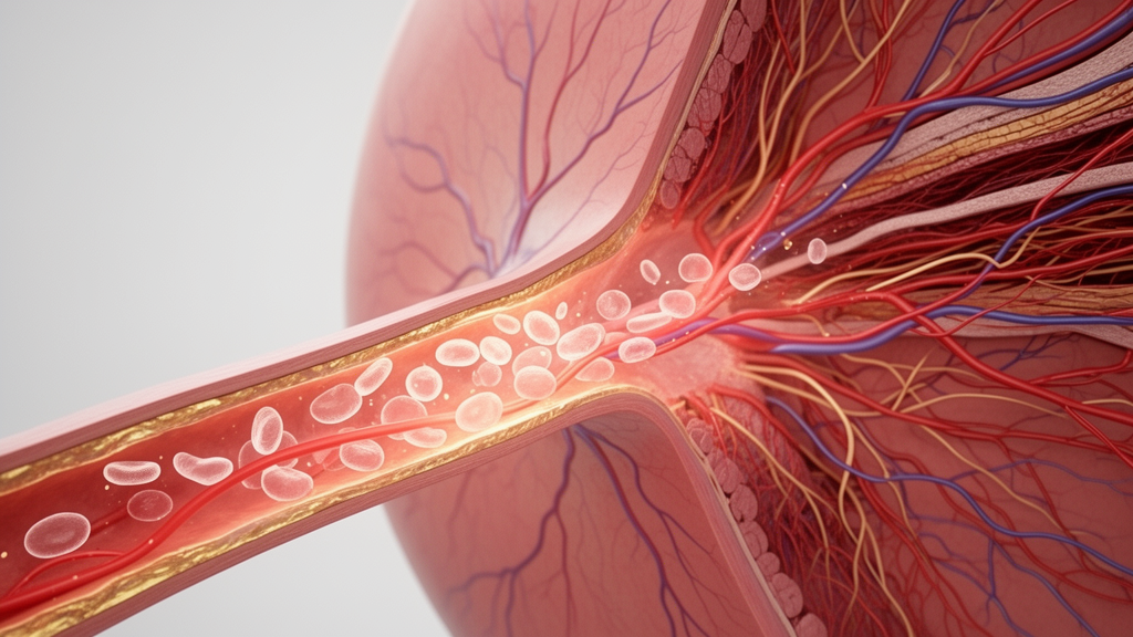
In a retrospective study of adults hospitalized with type 2 diabetes, people with more severe diabetic retinopathy showed lower brachial artery flow-mediated dilation (FMD) and larger platelets, two measures associated in the analysis with the eye disorder. Median FMD fell from 6.9% among participants without retinopathy to 6.1% in those with non-proliferative disease and 5.3% in those with proliferative disease, while median mean platelet volume (MPV) rose from 9.30 to 9.60 and 10.00 femtoliters, respectively. The findings are exploratory and do not show that either measurement predicts future retinopathy, causes it or has clinical value.
The hospital-based study analyzed routinely collected records from 109 adults with type 2 diabetes who completed a multidisciplinary assessment pathway at the First Affiliated Hospital of Sun Yat-sen University in Guangzhou between June 2015 and December 2019. Forty-six participants had no diabetic retinopathy, 36 had non-proliferative retinopathy and 27 had proliferative retinopathy. All received dilated fundus examinations and standardized fundus photography, and vascular ultrasound was performed irrespective of retinopathy status or cardiovascular risk profile. Because the data were collected during standard inpatient care rather than under a prospective research protocol, the analysis was cross-sectional and retrospective.
Two retinal specialists, blinded to clinical and ultrasound findings, graded each participant using the International Clinical Diabetic Retinopathy Disease Severity Scale. When the eyes differed, the participant was assigned the more severe grade. Because the numbers with mild, moderate and severe non-proliferative disease were too small for the intended statistical model, those categories were combined after the fact, reducing the ability to resolve differences within that stage. The investigators also examined carotid artery diameter, intima–media thickness and plaque presence as secondary outcomes. These comparisons were exploratory, and the group-wide statistical tests were not designed to test an ordered trend.
FMD is a noninvasive measure of endothelium-dependent vasodilation. During the examination, participants fasted and avoided caffeine, alcohol, smoking, vigorous exercise and vasoactive medication before resting supine for 15 minutes in a temperature-controlled room. An ultrasound operator measured the right brachial artery at rest, inflated a forearm cuff to 250 mm Hg for five minutes and recorded the artery after cuff release. FMD was calculated from the change between baseline and maximum post-deflation diameter relative to baseline. All examinations used the same equipment, protocol and experienced sonographer, but formal intraobserver repeatability was not assessed and post-deflation Doppler flow velocity was unavailable.
The main statistical model was a proportional-odds analysis treating retinopathy severity as an ordered outcome: no retinopathy, non-proliferative disease and proliferative disease. It simultaneously included FMD, MPV, age, sex, diabetes duration, glycated hemoglobin and systolic blood pressure. Of the 109 participants, 86 had complete data for this model—36 without retinopathy, 30 with non-proliferative disease and 20 with proliferative disease. The remaining 23 lacked MPV, glycated hemoglobin or both, and excluded participants had higher blood urea nitrogen and creatinine, raising the possibility of complete-case selection bias. The model was selected on clinical grounds rather than prospectively prespecified, and all estimates were treated as exploratory.
In that 86-person model, each one-percentage-point increase in FMD was associated with an adjusted odds ratio of 0.831 for greater retinopathy severity, with a 95% confidence interval of 0.713 to 0.968 and a P value of .018. In the opposite direction, each 1-femtoliter increase in MPV was associated with an adjusted odds ratio of 1.898, with a 95% confidence interval of 1.238 to 2.912 and a P value of .003. Diagnostic checks did not detect a violation of the proportional-odds assumption or a detectable nonlinear relationship among the continuous predictors, and leave-one-out refits retained the direction of both associations. Nevertheless, the modest sample, multiple analyses and lack of multiplicity adjustment make these results hypothesis-generating rather than confirmatory.
The associations changed when potential confounding and model stability were examined. Adding recorded diabetic nephropathy attenuated the FMD estimate to an odds ratio of 0.855, with a 95% confidence interval of 0.732 to 0.998, while the MPV estimate was preserved at 2.166, with a 95% confidence interval of 1.356 to 3.458. Renal status appeared more common as retinopathy advanced: recorded nephropathy affected 2.2% of participants without retinopathy, 16.7% with non-proliferative disease and 29.6% with proliferative disease. However, the analysis lacked participant-level estimated glomerular filtration rate and albuminuria, so the chart-based nephropathy variable may have misclassified renal disease and did not fully resolve confounding by kidney disease.
A ridge-penalized model offered a second warning about effect magnitude. At a penalty selected by repeated stratified cross-validation, the FMD odds ratio moved closer to the null, from 0.831 to 0.899 at the minimum-loss penalty and 0.919 at the one-standard-error penalty. The corresponding MPV estimates declined more sharply, from 1.898 to 1.388 and 1.289. Because penalized coefficients are shrunk toward zero by design, these figures are not corrected effect estimates; their attenuation nevertheless indicates that the unpenalized estimates may overstate the strength of the relationships in this small sample. Additional one-at-a-time adjustments for medication classes, carotid plaque, baseline brachial diameter and exclusion of the two participants with heart failure did not materially change the main estimates.
Secondary carotid findings were consistent with greater vascular abnormality among participants with more advanced eye disease, but they do not demonstrate that macrovascular changes caused retinopathy. Right carotid intima–media thickness had medians of 0.70, 0.80 and 0.89 millimeters across the three retinopathy categories, while any carotid plaque was present in 43.5%, 62.9% and 70.4%. Blood urea nitrogen and the triglyceride-to-HDL cholesterol ratio also differed across groups, while most major demographic, glycemic and blood-pressure comparisons did not show clear differences. These unadjusted group comparisons cannot establish temporal sequence and were vulnerable to confounding.
Several design features sharply limit interpretation. The hospital sample may not represent people with type 2 diabetes in the wider community, and indications for admission were not systematically recorded. The cross-sectional measurements cannot show whether lower FMD and larger platelets preceded retinal disease or instead reflected other complications, hospitalization-related changes or treatment. Eye imaging used one macula-centered 45-degree photograph per eye rather than widefield or multifield imaging, so peripheral lesions could have been missed. MPV was measured with one analyzer, making comparison within the study appropriate but limiting transfer of absolute values to other platforms. Because diabetic macular edema was documented for only 51 participants with highly uneven missingness, it could not be compared reliably.
The clearest conclusion is therefore narrow: in this selected group of hospitalized adults with type 2 diabetes, lower brachial FMD and higher MPV accompanied more severe diabetic retinopathy. The FMD relationship was sensitive to adjustment for recorded nephropathy, while both estimates weakened under penalization, especially the MPV association. The results do not establish causality, temporal sequence, predictive performance, diagnostic cutoffs or clinical utility. They instead provide a rationale for larger, prospective, multicenter cohorts to test whether endothelial function and platelet characteristics can help explain or anticipate the progression of diabetic eye disease.
Subject of Research: Cross-sectional associations of brachial FMD and mean platelet volume with diabetic retinopathy severity in adults with type 2 diabetes.
Article Title: Brachial artery flow-mediated dilation and mean platelet volume in relation to diabetic retinopathy severity in type 2 diabetes: a retrospective cross-sectional study
Article References: Zeng, Y., Wang, C., Huang, Z., Chen, T., Yao, F., & Zhu, W. (2026). Brachial artery flow-mediated dilation and mean platelet volume in relation to diabetic retinopathy severity in type 2 diabetes: a retrospective cross-sectional study. Frontiers in Endocrinology, 17, Article 1911136. https://doi.org/10.3389/fendo.2026.1911136
Image Credits: AI Generated
DOI: 10.3389/fendo.2026.1911136
Keywords: diabetic retinopathy, type 2 diabetes, flow-mediated dilation, mean platelet volume, endothelial function, platelet size, vascular ultrasound, retinopathy severity, cross-sectional study, Brachial, artery, flow-mediated
Cite Scienmag News
Reid Dalton. (October 23, 2026). Vascular and platelet markers track retinopathy severity in type 2 diabetes. Scienmag. https://scienmag.com/vascular-and-platelet-markers-track-retinopathy-severity-in-type-2-diabetes/
Reid Dalton. "Vascular and platelet markers track retinopathy severity in type 2 diabetes." Scienmag, 23 October 2026, https://scienmag.com/vascular-and-platelet-markers-track-retinopathy-severity-in-type-2-diabetes/. Accessed 23 October 2026.
Reid Dalton. "Vascular and platelet markers track retinopathy severity in type 2 diabetes." Scienmag. October 23, 2026. https://scienmag.com/vascular-and-platelet-markers-track-retinopathy-severity-in-type-2-diabetes/


