


Tuberculosis remains one of the world’s most stubborn infectious disease challenges, and the reason is not only the biology of the bacterium but also the sheer length of treatment. Curing drug-sensitive tuberculosis typically requires patients to take a combination of antibiotics for at least six months, and adherence over that long course is the single most important determinant of whether the infection is cleared, whether drug resistance emerges, and whether the disease spreads further in the community. Health systems have tried everything from directly observed therapy, in which a health worker watches each dose being swallowed, to pill counts, refill records, and digital pillboxes. Each approach carries costs, burdens, and blind spots. Now a research team reporting in PLOS Global Public Health has taken a careful, evidence-based look at a different strategy: a biochemical test that patients perform at home, photograph with a mobile phone, and upload through a companion app, allowing clinicians and treatment supporters to see objective evidence of recent medication ingestion.
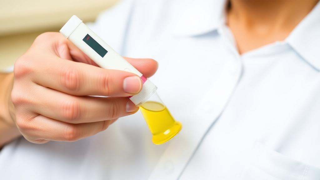
The technology at the heart of the study is called the TB Treatment Support Tools, or TB-TST, system. It pairs a urine-based lateral flow assay with a mobile application designed for patient education, encouragement, and communication with treatment supporters. The lateral flow format will be familiar to anyone who has used a pregnancy test or a rapid antigen test: a sample wicks along a strip, and colored lines appear where target molecules are captured. In this case, the target is not the tuberculosis bacterium itself but a metabolite of isoniazid, one of the cornerstone drugs of TB therapy. When a patient takes isoniazid, the body breaks the drug down and excretes its metabolic products in urine. The test strip detects these metabolites, and a blue-purple color change signals that the medication was ingested recently. A negative result, in principle, means no recent dose was taken, giving caregivers a biochemical window into adherence that self-reporting simply cannot provide.
Because the test is read from a photograph rather than by a trained technician standing in a clinic, the entire approach depends on patients being able to capture usable images and on those images being interpretable at a distance. That is the question the research team set out to answer. In a pragmatic randomized controlled trial, participants aged sixteen or older who had been newly diagnosed with tuberculosis and had access to a mobile phone used the system as part of their routine care, uploading photographs of their test cartridges through the app. Rather than relying on the impressions of individual readers, the investigators applied a structured qualitative method known as visual content analysis, systematically coding 1,655 user-submitted test images to identify recurring patterns and problems that affected interpretation.
The coding process was iterative, meaning the researchers developed and refined a codebook as they reviewed images, adding and sharpening categories until they captured the full range of what appeared on screen. Three primary issue categories emerged from this analysis. The first concerned color variations in the test and control strips, which is critical because the assay’s meaning hinges on recognizing the correct blue-purple signal. The second category covered photo quality issues, since a blurry, poorly lit, or badly framed image can obscure even a perfectly executed test. The third category involved artifacts of the test cartridge and strip themselves, physical quirks of the device that can mimic or distort the expected color patterns.
The headline finding is reassuring for the technology’s prospects. Most of the submitted images, 72 percent, were consistent with either a clear positive result showing the characteristic blue-purple color change or a clear negative. In other words, despite the fact that the photographs were taken by patients in real-world home settings, with all the variability in lighting, skill, and device quality that implies, the large majority of tests could be read with confidence. That proportion is a meaningful benchmark for any home-based diagnostic or monitoring tool, and it suggests that the burden of interpretation does not fall disproportionately on ambiguous results that would force clinicians to guess.
The remaining images, however, reveal exactly where the system’s weak points lie, and the researchers cataloged them in detail. Among the results that were not clear positives, the team observed strips showing no color change at all, appearing white; strips displaying yellow or orange hues; and strips with green or teal coloration. Each of these atypical outcomes raises interpretive questions. A white strip could reflect a failed test or an expired cartridge. Yellow and orange tones might represent intermediate chemistry on the way to the expected color change, while green and teal hues fall outside the expected palette entirely. Without clear protocols for handling these atypical colors, readers may hesitate, misclassify, or request repeat tests, all of which erode the efficiency that home-based monitoring is supposed to deliver.
Photo quality proved to be the second major source of difficulty, and blurriness was by far the most common image quality issue encountered. This is an intuitive but important finding: smartphone cameras are capable of excellent macro photography, but patients holding a small plastic cartridge at arm’s length, perhaps in imperfect lighting, do not always achieve focus. The good news embedded in this finding is that image quality is arguably the most tractable of the problems identified. Unlike the chemistry of the assay, which is fixed by the test’s design, the quality of a photograph can be improved through user guidance, in-app prompts, automatic focus checks, and real-time feedback that tells a patient before submission whether the image is sharp enough to read.
The third category, test cartridge and strip artifacts, points to engineering rather than behavioral fixes. The most frequently observed artifact was visual evidence consistent with dye bleed, or possible cross-strip contamination, between the control and test lines of the device. In a lateral flow assay, the control line confirms that the test functioned properly, while the test line carries the diagnostic signal. When dye migrates or bleeds from one region to another, the boundary between those signals can blur, complicating the reading. This kind of artifact is a design problem, and the authors argue that refinements to the cartridge itself could reduce its occurrence, protecting the clarity of results at the source rather than compensating for it downstream.
What makes this study notable methodologically is its fusion of qualitative image analysis with engineering design thinking. Instead of treating ambiguous images as noise to be discarded, the team treated them as data about the user experience, each problematic photograph a clue about where the system, the instructions, the device, or the interpretation protocol was failing the patient. This user-centered framing transforms a pile of blurry and oddly colored pictures into a concrete improvement roadmap. The authors identify four priority directions: enhanced user guidance for image capture, refinement of cartridge design to reduce artifacts, clearer interpretation protocols for atypical color outcomes, and automated image-based reading, in which software rather than a human eye would classify the strip, potentially standardizing results and reducing reader burden simultaneously.
The broader implications extend well beyond tuberculosis. Digital adherence technologies are a growing field, and pairing biochemical verification with mobile image reporting is an elegant way to combine objectivity with patient autonomy, allowing people to test at home while still generating verifiable evidence for their care teams. The finding that relatively few images were truly difficult to interpret, despite the challenges in submission and test appearance, supports the feasibility of user-submitted lateral flow images for monitoring recent isoniazid ingestion in routine care. As home-based testing expands across infectious disease, chronic disease management, and beyond, the lessons from this trial are clear: the chemistry of a test is only half the product, and the other half is the human being holding the phone. Designing for that person, with better guidance, better hardware, and smarter automated reading, is what will turn a promising prototype into a dependable public health tool.
Subject of Research: Evaluation of a urine-based lateral flow assay for monitoring tuberculosis medication adherence through patient-submitted smartphone images
Article Title: Evaluating a biochemical TB medication adherence test: Insights from image-based reporting through a patient companion app
Article References: Lee, K., Liang, E. C., Tolentino, A., Trinh, A., Leon, D., Goodwin, K., Sprecher, J., Rubinstein, F., Lutz, B., & Iribarren, S. (2026). Evaluating a biochemical TB medication adherence test: Insights from image-based reporting through a patient companion app. PLOS Global Public Health, 6(10), e0007182. https://doi.org/10.1371/journal.pgph.0007182
Image Credits: AI Generated
DOI: 10.1371/journal.pgph.0007182
Keywords: tuberculosis, medication adherence, lateral flow assay, isoniazid metabolite, mobile health, digital adherence technology, urine test, image analysis, patient companion app, randomized controlled trial, PLOS Global Public Health, diagnostic usability
Cite Scienmag News
Ophelia Keating. (October 10, 2026). Urine Test Photos Sent by TB Patients Prove Reliable for Checking Medication Intake. Scienmag. https://scienmag.com/urine-test-photos-sent-by-tb-patients-prove-reliable-for-checking-medication-intake/
Ophelia Keating. "Urine Test Photos Sent by TB Patients Prove Reliable for Checking Medication Intake." Scienmag, 10 October 2026, https://scienmag.com/urine-test-photos-sent-by-tb-patients-prove-reliable-for-checking-medication-intake/. Accessed 10 October 2026.
Ophelia Keating. "Urine Test Photos Sent by TB Patients Prove Reliable for Checking Medication Intake." Scienmag. October 10, 2026. https://scienmag.com/urine-test-photos-sent-by-tb-patients-prove-reliable-for-checking-medication-intake/


