In the rapidly evolving landscape of medical imaging and artificial intelligence, a groundbreaking development has recently emerged that promises to revolutionize how clinicians interpret full-body scans and enhance diagnostic accuracy. A research group led by Chen, Y., Gao, L., and colleagues has unveiled an innovative modality-projection universal model aimed at full-body medical imaging segmentation—a formidable challenge given the diversity and complexity of imaging modalities and anatomical variations. This new approach, elaborated in their seminal paper published in Nature Communications, introduces a conceptually elegant yet technologically sophisticated framework for seamless segmentation across multiple imaging modalities.
The crux of the problem addressed by Chen et al. lies in the intrinsic disparity between varied medical imaging types such as computed tomography (CT), magnetic resonance imaging (MRI), and positron emission tomography (PET). Traditionally, segmentation techniques have been modality-specific, crafted and honed separately for each imaging type due to differences in image characteristics, signal intensity profiles, and noise patterns. This fragmentation hampers comprehensive cross-modality image analysis and often impedes the integration of multi-modal datasets critical for holistic patient evaluations. Recognizing this limitation, the researchers developed a universal model that successfully projects and unifies these heterogeneous data types into a shared representational space, facilitating accurate and consistent segmentation results.
Central to the proposed method is the notion of modality projection, whereby multi-modal images are transformed and aligned via deep learning architectures capable of capturing modality-invariant features. This technique counters the traditional paradigm of training separate models, instead relying on a shared latent space representation that enables simultaneous processing. By leveraging convolutional neural networks enriched with spatial and contextual awareness, the model effectively disentangles modality-specific visual cues from underlying anatomical structures, thereby preserving the essential features needed for precise segmentation irrespective of input imaging modality.
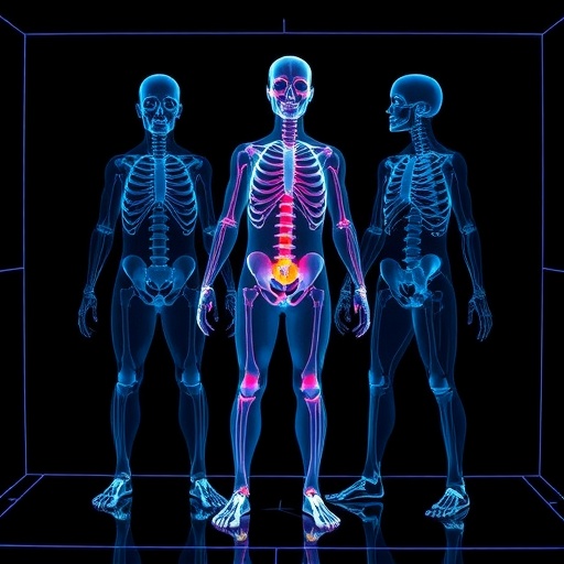
In testing their model on an extensive collection of full-body images from various modalities, the researchers demonstrated superior segmentation accuracy and generalizability compared to existing state-of-the-art methods. Notably, the framework was adept at delineating critical anatomical regions across the entire body, including complex structures such as vascular networks, skeletal features, and soft tissues. This wide anatomical coverage is unprecedented in the field, moving beyond the common focus on discrete organs or regions of interest. The ability to segment full-body scans with such granularity and reliability opens new frontiers for clinical applications, from advanced diagnostics and surgical planning to personalized medicine.
Integral to the model’s success is its innovative architecture, meticulously designed to incorporate modality-specific encoders followed by a shared decoder pathway. This structural design ingeniously balances the need to extract unique modality features while converging on a universal segmentation output. The encoders function to preprocess and standardize each modality, effectively normalizing signal discrepancies. Subsequently, the shared decoder leverages a unifying feature space to output segmentation maps that maintain consistency across modalities. This architectural insight marks a paradigm shift in medical image analysis, offering a scalable solution adaptable to future imaging technologies.
Furthermore, the model integrates robust attention mechanisms that dynamically weigh the importance of features extracted from different modalities, enhancing interpretability while boosting performance. These attention layers allow the system to prioritize salient anatomical information based on clinical context and image quality, thus mitigating noise and artifacts inherent to certain imaging techniques. The dynamic feature weighting enhances the overall fidelity of the segmentation and provides clinicians with reliable and interpretable outputs critical for decision-making processes.
Beyond technical achievements, the model also emphasizes clinical usability and integration. Chen and colleagues implemented an intuitive user interface compatible with existing radiological imaging workflows, facilitating seamless adoption by healthcare professionals. Real-time processing speed and scalability underpin the model’s practical deployment potential, ensuring that the technology can be incorporated into busy clinical environments without compromising throughput. The interoperability with hospital information systems signals a promising trajectory towards routine clinical implementation.
Another transformative aspect of this study is its contribution to tackling challenges faced in rare and complex diseases where multi-modal imaging often conveys complementary pathological insights. By enabling cohesive segmentation across diverse scanning techniques, the model aids in comprehensive disease characterization and monitoring, which is crucial for conditions involving multisystem involvement such as systemic lupus erythematosus or metastatic cancers. The advancement portends improved patient stratification, prognosis estimation, and tailored therapeutic interventions.
The ramifications of this innovation extend into research domains as well. Multi-center clinical trials often grapple with heterogeneous imaging data due to protocol differences or equipment variability. A universal segmentation model such as this could harmonize imaging datasets, allowing for more robust cross-study comparisons and meta-analyses. Researchers gain the ability to pool data with greater confidence in image-derived biomarkers, accelerating biomarker discovery and validation processes essential for translational medicine.
Importantly, the model’s training regime incorporates sophisticated data augmentation and domain adaptation strategies to mitigate overfitting and enhance generalizability across patient demographics and scanner types. These methodological refinements ensure that the model performs reliably across populations, imaging hardware, and clinical settings, a pivotal advantage that addresses the often-cited challenge of AI bias in medical imaging. The emphasis on model robustness reflects growing awareness of the necessity for equitable AI applications in healthcare.
From a technical perspective, the deployment of extensive pre-training on large, annotated datasets coupled with fine-tuning on institution-specific data sets a new standard in model optimization. This hybrid strategy optimizes the model’s ability to leverage generalized anatomical knowledge while adapting to specific clinical environments. The approach exemplifies a judicious balance between data efficiency and performance, mitigating common bottlenecks such as limited annotated medical data availability.
The implications for future research trajectories are profound. This universal model could serve as a foundational platform upon which specialized downstream segmentation tasks can be built, allowing for rapid customization and extension. Researchers may develop plug-in modules addressing particular clinical needs or pathologies, thereby fostering an ecosystem of interoperable AI tools enhancing the versatility and scalability of imaging workflows.
Moreover, the ethical dimension of deploying such powerful AI tools was addressed with careful consideration by the authors. Their framework incorporates transparency measures and uncertainty quantification, vital for clinician trust and regulatory compliance. The model’s interpretability features support the explainability crucial in high-stakes medical decisions, ensuring that augmented intelligence complements human expertise responsibly.
Anticipated future iterations of this technology envision integration with other diagnostic modalities such as genomics and laboratory data, moving towards a truly multi-omics precision medicine paradigm. This integrative approach promises to bridge gaps between imaging phenotypes and molecular profiles, unlocking deeper insights into disease etiology and progression.
In summary, this pioneering universal modality-projection model represents a milestone in medical image segmentation, harnessing advanced deep learning techniques to unify diverse imaging modalities into a coherent analytical framework. Its blend of high accuracy, scalability, and clinical pragmatism augurs well for accelerated adoption and transformative impacts on diagnostic medicine. As healthcare increasingly embraces AI-driven precision, such integrative models will serve as cornerstones for the next generation of comprehensive, patient-centric medical imaging solutions.
Subject of Research: Universal deep learning model development for full-body medical imaging segmentation across multiple modalities.
Article Title: Modality-projection universal model for comprehensive full-body medical imaging segmentation.
Article References:
Chen, Y., Gao, L., Gao, Y. et al. Modality-projection universal model for comprehensive full-body medical imaging segmentation. Nat Commun 16, 9423 (2025). https://doi.org/10.1038/s41467-025-64469-w
Image Credits: AI Generated
