


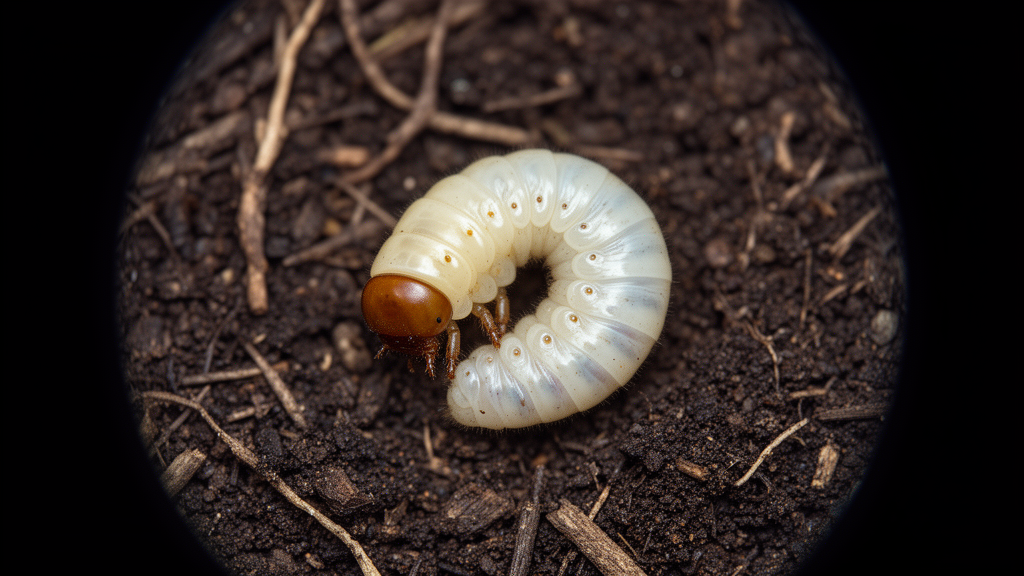
Deep in the forests of northeastern China, a small beetle has been hiding its most intimate secrets. Now, for the first time, scientists have pulled back the curtain on the immature stages of Parastasia ferrieri, a member of the shining leaf chafers, using both light and scanning electron microscopy to reveal a larva unlike most of its relatives. The findings, published in the Deutsche Entomologische Zeitschrift, offer a rare glimpse into the hidden world of white grubs and may help researchers understand how these beetles fit into forest ecosystems.
Shining leaf chafers, the subfamily Rutelinae, get their name from the bright, often metallic colors of their adults, which feed on plant leaves. The group is enormous: more than 4,000 species distributed among 235 genera, organized into seven tribes. Yet despite the dazzling adults, the larvae of these beetles, commonly called white grubs, remain largely unknown. Fewer than 2 percent of all known scarab species have had their larvae described, a gap that frustrates ecologists who cannot reliably identify which grub belongs to which beetle, and therefore cannot generalize the ecological role of any given species until its immature stages have been accurately matched.
This is where Parastasia ferrieri comes in. Rutelini, the tribe to which it belongs, encompasses roughly a thousand species in 93 genera, with a primary presence in the New World. Recently, both larvae and adults of P. ferrieri were discovered in Liaoning Province of northeastern China, in the Palearctic realm, similar to previous records in Korea. Parastasia is one of the largest genera within Rutelini, comprising approximately 105 species worldwide, but unlike many of its showy relatives, its adults lack enlarged mandibles, thoracic horns, or vibrant metallic coloration. That modesty may explain why the species has gone largely unnoticed.
The research team collected 13 larvae from Qipanshan Forest Park in Shenyang City in late October 2019. The larvae were reared in plastic boxes filled with moist, fermented sawdust, with a decayed wood log off-cut provided to facilitate boring and egg-laying. Adults emerged the following May, and third instar larvae were collected from the sawdust in September. Ten larvae were fixed in Dietrich’s solution, heated to 70 degrees Celsius and left to cool naturally for 12 hours before preservation in 75 percent ethanol. For scanning electron microscopy, dissected organs underwent serial dehydration in graded ethanol, replacement with tertiary butanol, freeze-drying for 3 hours, and sputter-coating with gold before examination under a Hitachi S-3400N scanning electron microscope at 5 kV.
The larvae are of typically scarabaeiform shape, bearing three pairs of thoracic legs on a C-shaped body. The trunk is generally white, with a yellowish head capsule, thoracic legs, and spiracles. The head capsules measure 3.1 millimeters in width on average and display symmetrical adornment with a total of 14 pairs of setae: two pairs on the clypeus, five pairs in the frontal region, three pairs on the vertex, and four pairs in the genal area. The antenna is elongated and slender, comprising four segments with the second being the longest, and the distal conical segment bears seven sensilla basiconica at its apex.
The mouthparts are of a biting-chewing type, and it is here that the larva reveals its most remarkable features. The labrum, slightly wider than it is long, displays four prominent protuberances on its outer surface, an atypical trait not mentioned in the congeneric larvae of P. brevipes. The epipharynx, the membranous structure on the inner surfaces of the labrum and clypeus, is divided into distinct functional areas including a haptomerum at the apex, a pair of plegmatia on the lateral margins, and glabrous gymnoparia on both sides. Notably, the haptomerum lacks a helus, consistent with previous descriptions in Rutelini, where heli are entirely absent, in contrast to Anomalini with two to four, Anoplognathini with six to eight, and Adoretini with six to nine.
The paired mandibles are heavily sclerotized and symmetric, each bearing two scissorial teeth with apical teeth curved inward. This symmetry is unusual within Rutelini, where larval mandibles are typically asymmetric, equipped with three teeth on the left and two on the right. Each mandible also bears a shuttle-shaped stridulatory area on its ventral surface. The molar region is asymmetric, featuring a prominent molar tooth with an acia on the left and a group of four ridged molar teeth on the right. The maxillae, labia, and hypopharynx are fused into a structural complex, with each maxilla bearing a row of five stridulatory teeth and an anterior truncate process, and the maxillary palp consisting of four segments with ten sensilla basiconica on its apex.
The thoracic legs are composed of five segments each: coxa, trochanter, femur, tibiotarsus, and a distal single claw. The claw is heavily sclerotized with an obtuse distal end, congruent with other saproxylic larvae in Lucanidae, the wood-decomposing beetles. In most Melolonthinae and Cetoniinae, the claw is typically curved, sharp, and adorned with paired setae, while in Rutelinae, thoracic claws are typically sharp in Adoretini, Geniatini, and Anomalini. The blunt claws of P. ferrieri, combined with its other features, point toward a saproxylic lifestyle, feeding on decaying wood rather than living roots.
This correlation between morphology and feeding habits is central to the study’s discussion. Mandibles, being the most heavily sclerotized structures on the mouthparts, are structurally correlated with feeding habits across scarab lineages. Straight and apically sharp incisors appear in wood-consuming species of Lucanidae and Passalidae, sharp but inwardly curved incisors in dung decomposers within Aphodiinae and Scarabaeinae, and blade-like cutting edges in phytophagous Melolonthinae. Within Rutelinae, mandibular incisors are typically blade-like in phytophagous larvae of Anomalini and Adoretini, and apically sharp and curved in saproxylic larvae of Rutelini. The larvae of P. ferrieri, with their symmetric two-toothed mandibles and blunt claws, fit the saproxylic pattern seen in the previously described P. brevipes, which is reported to feed on dead wood.
The significance of this work extends beyond taxonomy. In the Palearctic realm, particularly in northeastern China, larvae of Rutelinae are frequently recognized as agricultural pests because they mostly belong to the phytophagous Anomalini or Adoretini, attacking living roots of crops and trees. The scarcity of records of P. ferrieri may be attributed to their small, nocturnal adults, or the fact that their larvae never attack living organs of crops or trees. Given their biogeographical significance, the researchers argue that the larvae of P. ferrieri warrant increased attention for conservation purposes. By documenting the larval morphology of this Palearctic Rutelini in such detail, the study adds a crucial piece to the puzzle of scarab diversity, helping researchers distinguish saproxylic decomposers from root-feeding pests and ultimately understand the ecological roles these hidden grubs play in forest ecosystems.
Subject of Research: Larval morphology and feeding habits of the Palearctic ruteline beetle Parastasia ferrieri
Article Title: Larval morphology of a Palearctic Rutelini , Parastasia ferrieri ( Coleoptera , Scarabaeidae ), with discussions on their feeding habits
Article References: Sun, X.-Y., Dong, X.-M., & Jiang, L. (2024). Larval morphology of a Palearctic Rutelini, Parastasia ferrieri (Coleoptera, Scarabaeidae), with discussions on their feeding habits. Deutsche Entomologische Zeitschrift, 71(1), 185-192. https://doi.org/10.3897/dez.71.113727
Image Credits: AI Generated
Keywords: Parastasia ferrieri, Rutelini, Rutelinae, Scarabaeidae, larval morphology, scanning electron microscopy, white grubs, saproxylic, feeding habits, Palearctic, China, entomology
Cite Scienmag News
Gavin Prescott. (October 19, 2026). Under the microscope: the secret larva of a shining leaf chafer. Scienmag. https://scienmag.com/under-the-microscope-the-secret-larva-of-a-shining-leaf-chafer/
Gavin Prescott. "Under the microscope: the secret larva of a shining leaf chafer." Scienmag, 19 October 2026, https://scienmag.com/under-the-microscope-the-secret-larva-of-a-shining-leaf-chafer/. Accessed 19 October 2026.
Gavin Prescott. "Under the microscope: the secret larva of a shining leaf chafer." Scienmag. October 19, 2026. https://scienmag.com/under-the-microscope-the-secret-larva-of-a-shining-leaf-chafer/


