


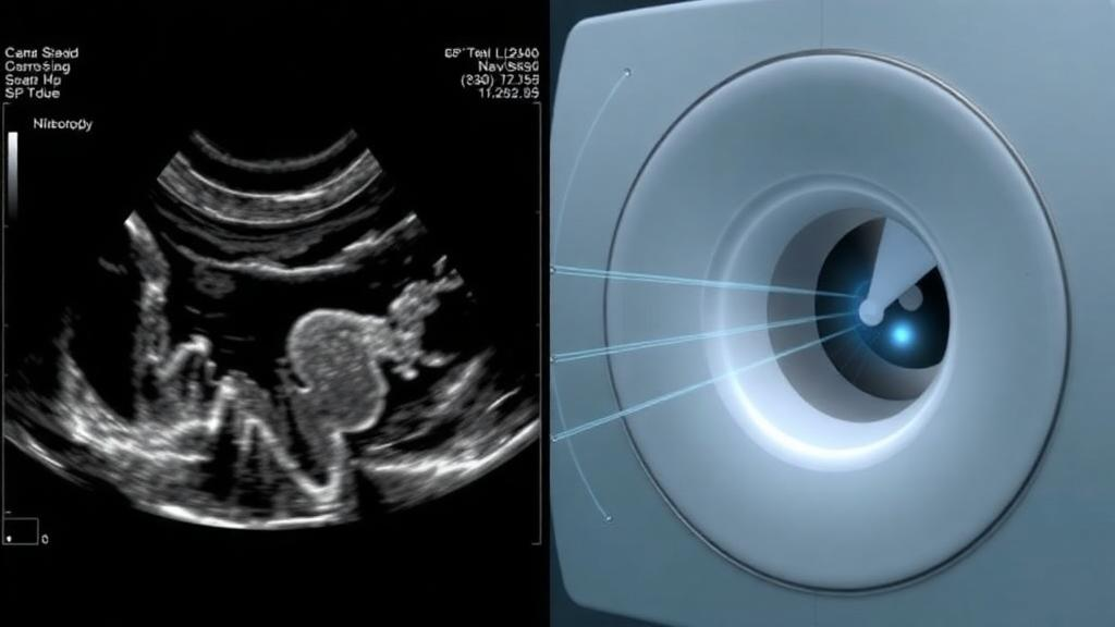
Spine surgery is an exercise in millimeters. A pedicle screw placed just two or three millimeters off its intended trajectory can irritate a nerve root, breach a vessel, or fail to hold the bone it is meant to stabilize. Surgeons have long relied on intraoperative imaging to guide their instruments, but every X-ray-based technique adds radiation dose to both patient and operating team. A new study from Beijing Shijitan Hospital, Capital Medical University, published in BMC Medical Imaging, offers a carefully quantified step toward a lower-dose alternative: fusing live, radiation-free ultrasound images with three-dimensional C-arm cone-beam computed tomography, and proving in a controlled laboratory setting that the resulting fusion is accurate to roughly one millimeter.
The research, led by Zerui Sun and Yingdong Hu with senior author Hongxing Song, tackles a deceptively hard engineering problem. Cone-beam CT, acquired by rotating a mobile C-arm around the patient, delivers an excellent three-dimensional map of bony anatomy, but it is a snapshot that quickly goes stale as the body moves. Tracked ultrasound, by contrast, can be swept across the spine in real time and carries no ionizing radiation, yet its images are noisy, prone to artifacts, and often show only fragments of the bone surface. Stitching the two modalities together requires knowing exactly where the ultrasound probe is in space, exactly how the ultrasound image relates to the probe, and how to mathematically align a partial, artifact-laden surface to a complete CT-derived bone model. Each step introduces error, and the study’s central contribution is measuring, with unusual rigor, how much error each step actually contributes.
The team built an integrated system combining an ultrasound machine, a C-arm capable of cone-beam CT, an optical tracking system that follows instruments in space, a robot-controlled probe holder that sweeps the ultrasound transducer at a constant speed, and a navigation workstation that fuses everything together. Rather than testing on patients, they used a lumbar spine phantom covering the L1 to L4 vertebrae, scanned while submerged in water. Water eliminates the air-tissue interfaces that plague ultrasound in real bodies, giving the researchers a best-case environment in which to isolate the performance of the registration mathematics themselves. That choice matters for interpreting the results: this is a validation of a workflow, not yet a demonstration in living tissue.
The first technical hurdle was probe calibration, the process of determining the precise transformation between the ultrasound image pixels and the tracked probe coordinate frame. Getting this wrong by even a fraction of a millimeter poisons every downstream measurement. The researchers designed an optimized double-layer phantom containing four N-shaped wires, a classic calibration geometry in which wires cross the imaging plane at known orientations, and calibrated the probe using eighty image sets. The ultrasound image scale came out to 0.16 millimeters per pixel. The full-data calibration error, computed as a pooled three-dimensional vector root-mean-square over all residuals, was 0.627 millimeters, and the per-image-set RMS across the eighty sets averaged 0.621 plus or minus 0.086 millimeters. Critically, the team also performed a split-half held-out validation, calibrating on one subset of images and testing on a disjoint subset, and the error barely moved, to 0.639 plus or minus 0.052 millimeters. That held-out figure is the more honest number, because it shows the calibration generalizes rather than merely fitting the data it was tuned on.
With calibration established, the harder problem was registration: aligning the ultrasound-derived bone surface to the cone-beam CT surface. The CBCT volume was segmented using a 400-Hounsfield-unit threshold with manual refinement, a standard approach for isolating cortical bone. The researchers then applied a three-stage registration pipeline. A similarity transform provided a coarse initial alignment, a multiresolution point-to-point iterative closest point algorithm, or ICP, refined the rigid alignment across progressively finer scales, and finally a thin-plate spline, or TPS, deformation absorbed the residual nonrigid mismatch between the two surfaces. The thin-plate spline is a mathematically elegant interpolant, often described as the higher-dimensional analogue of a flexible metal sheet bent over fixed control points, and it allowed the ultrasound surface to warp slightly to accommodate soft-tissue artifacts and incomplete visibility without abandoning the rigid skeleton of the alignment.
The headline numbers are striking. Evaluated on ten independently acquired ultrasound scans at eight bilateral pedicle-center landmarks that had been deliberately withheld from the optimization process, the point-to-point ICP stage alone achieved a mean target registration error of 2.98 plus or minus 0.23 millimeters. After the thin-plate spline refinement, that error dropped to 1.26 plus or minus 0.08 millimeters, a statistically significant improvement with a p-value below 0.001. Whole-surface distance metrics told the same story: the mean, RMS, and 95th-percentile Hausdorff distances fell from 1.80, 2.05, and 3.61 millimeters to 0.77, 0.88, and 1.54 millimeters respectively. In practical terms, the fused ultrasound view of the spine matched the CT model to within roughly the thickness of a coin at the exact landmarks where a surgeon would place pedicle screws.
One of the study’s most valuable findings is almost a negative result. The team compared point-to-point ICP with point-to-plane ICP, a variant that aligns surface normals and often converges faster and more accurately on rigid surfaces. Point-to-plane did modestly improve the rigid registration stage, but when the full pipeline including the TPS refinement was run, the final target registration error changed by only 0.005 millimeters. The lesson is that the nonrigid refinement stage, not the choice of rigid ICP formulation, is what drives the final accuracy. Supplementary computational analyses probing operator effects, scan pitch, frame density, and dynamic inconsistency reinforced the picture that the pipeline is robust to reasonable variations in acquisition conditions.
Speed matters as much as accuracy in an operating room, and the timing figures are sobering but transparent. The effective ultrasound acquisition region spanned roughly 170 millimeters, which at a sweep speed of 5 millimeters per second takes about 34 seconds of robot-controlled probe motion. The cumulative machine-processing time after scanning was 143.7 seconds, though the thin-plate spline solve itself required only about 0.06 seconds. The authors are explicit that this human-in-the-loop workflow is not suitable for continuous real-time use; it is a registration procedure performed once per case, not a live-updating navigation display. For procedures like pedicle screw placement, where anatomy is fixed relative to the navigation frame once the patient is positioned, a one-time registration of this accuracy could still be clinically transformative, cutting radiation exposure while preserving three-dimensional guidance.
The authors are equally candid about the limits of what they have shown. Because the study used a water-submerged phantom, the notorious degradation of ultrasound in real tissue, with its shadowing from bone, scattering from fat, and air artifacts, remains untested. No cadaveric, animal, or clinical validation has yet been performed, and the study explicitly does not validate a complete robot-assisted surgical system because instrument execution, needle insertion, and robotic puncture were not assessed. The dynamic robustness analyses were computational rather than physical. These caveats are not footnotes; they are the difference between a promising engineering result and a proven clinical tool.
Still, the significance of the work lies in its methodological discipline. By withholding landmark points from optimization, reporting held-out calibration errors, using run-level statistics across ten independent scans, and quantifying every stage of the pipeline separately, the Beijing team has produced one of the more rigorous accuracy characterizations of ultrasound-to-CBCT fusion in the spine navigation literature. Millimeter-level fusion accuracy, achieved without any additional patient radiation beyond a single low-dose cone-beam acquisition, is exactly the performance envelope that next-generation navigation systems will need if ultrasound is to take on a larger role in the operating room. The road from phantom to patient runs through tissue, cadavers, and clinical trials, but this study has established a credible, precisely measured starting point for that journey.
Subject of Research: Phantom-based accuracy validation of ultrasound and C-arm cone-beam CT image fusion for spine navigation
Article Title: Phantom-based accuracy validation of multimodal ultrasound and C-arm cone-beam CT image fusion for spine navigation
Article References: Sun, Z., Hu, Y., Fan, Y., Wang, J., & Song, H. (2026). Phantom-based accuracy validation of multimodal ultrasound and C-arm cone-beam CT image fusion for spine navigation. BMC Medical Imaging. https://doi.org/10.1186/s12880-026-02889-w
Image Credits: AI Generated
DOI: 10.1186/s12880-026-02889-w
Keywords: ultrasound, cone-beam CT, image fusion, spine navigation, target registration error, phantom study, probe calibration, iterative closest point, thin-plate spline, surgical navigation, radiation dose reduction, medical imaging
Cite Scienmag News
Ophelia Keating. (October 12, 2026). Ultrasound Meets Cone-Beam CT: Phantom Test Hits Millimeter Precision for Spine Surgery Navigation. Scienmag. https://scienmag.com/ultrasound-meets-cone-beam-ct-phantom-test-hits-millimeter-precision-for-spine-surgery-navigation/
Ophelia Keating. "Ultrasound Meets Cone-Beam CT: Phantom Test Hits Millimeter Precision for Spine Surgery Navigation." Scienmag, 12 October 2026, https://scienmag.com/ultrasound-meets-cone-beam-ct-phantom-test-hits-millimeter-precision-for-spine-surgery-navigation/. Accessed 12 October 2026.
Ophelia Keating. "Ultrasound Meets Cone-Beam CT: Phantom Test Hits Millimeter Precision for Spine Surgery Navigation." Scienmag. October 12, 2026. https://scienmag.com/ultrasound-meets-cone-beam-ct-phantom-test-hits-millimeter-precision-for-spine-surgery-navigation/


