


Every year, thousands of young children tumble from playground equipment, beds, and living room furniture, landing on an outstretched arm and fracturing the bone just above the elbow. Known as supracondylar humerus fractures, these injuries are the most common elbow fractures in childhood, and they demand precise surgical repair. Now, a team of orthopedic surgeons in China reports that a deceptively simple twist of a standard surgical wire may make that repair safer, reporting zero pin-related complications in a group of sixty children treated with their modified technique.
The study, published in BMC Pediatrics by Xiangyu Lv, Bicheng Liu, Yu Wang, Chenglei Zhao, and Jingxin Zhao of the Affiliated Hospital of Chengde Medical University, examined a modification of the classic Kirschner wire fixation procedure. Kirschner wires, often called K-wires, are thin, smooth steel pins that surgeons drill through bone to hold fractured fragments in place while the body heals. In pediatric elbow fractures, the standard operation, closed reduction and percutaneous pinning, involves manipulating the broken bone back into alignment without opening the joint and then threading pins through the skin to lock the fragments together. It is an elegant, minimally invasive approach, but it carries a well-known weakness: smooth pins can slide.
That weakness is not trivial. Because K-wires are deliberately smooth to minimize tissue damage during insertion and removal, they rely entirely on friction and geometry to stay put. In young children, whose bones are soft and whose casts and cooperation are less reliable than an adult’s, pins can migrate within the bone, back out through the skin, or lose their grip on the fracture fragments altogether. Pin migration can lead to loss of fracture alignment, skin irritation and infection at the pin site, and occasionally the need for unplanned revision surgery. Surgeons have long sought ways to anchor the pins more securely without adding operative time, cost, or iatrogenic injury, meaning harm caused by the treatment itself.
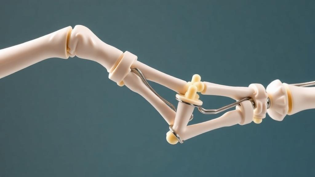
The modification tested by the Chengde team addresses the problem at the outer end of the wire rather than inside the bone. In the standard technique, two K-wires are inserted from the lateral humeral condyle, the bony prominence on the outer side of the elbow, crossing the fracture site and engaging the cortex, the dense outer layer of bone, on the opposite side of the humerus. This lateral divergent configuration is widely used because it avoids the ulnar nerve, which runs along the inner elbow and is at risk when pins are placed from the medial side. The Chengde group kept this configuration but added a final step: after both wires were seated, the surgeons rotated the exposed external ends of the wires and tightened them, securing the pins against each other and against back-out.
To test whether this extra maneuver mattered, the researchers conducted a retrospective comparative study of 120 children under eight years old with supracondylar humerus fractures, all treated at their hospital between January 2016 and June 2024. All eligible patients during the study period were consecutively included, with no random sampling. Sixty children received the modified technique and sixty received the standard two-wire fixation without the rotational step. The team compared operative time, time to fracture union, elbow function assessed with Flynn’s criteria, a widely used scoring system for pediatric elbow outcomes, and the occurrence of complications such as wire migration and fixation failure.
The headline finding was striking, though the authors are careful about its statistical weight. In the modified-technique group, there were no cases of K-wire migration within the bone. In the control group, four children, or 6.7 percent, experienced migration. The overall complication rate was zero percent in the study group compared with 8.33 percent in the control group. However, with sixty patients per arm, neither difference reached conventional statistical significance; the migration comparison yielded a p-value of 0.119 and the overall complication comparison a p-value of 0.057, both calculated with Fisher’s exact test, which is appropriate for small counts in contingency tables. The authors describe the technique as feasible and effective, noting that it partially overcomes the limitations of smooth K-wires while mitigating iatrogenic injury, and they frame the results as supporting clinical utility and potential for broader application rather than as definitive proof of superiority.
That caution is scientifically appropriate, and it highlights an important lesson in how to read surgical studies. A p-value of 0.057 sits just beyond the conventional 0.05 threshold, and in a study of this size, an observed difference of eight percentage points in complications cannot be distinguished with confidence from the play of chance. Absence of statistical significance is not evidence of no effect; it is evidence that the study was not large enough to settle the question. A randomized trial with more patients would be needed to confirm that the rotational tightening step genuinely reduces complications. The retrospective design also introduces the possibility of selection effects, since patients were grouped by the technique they actually received rather than randomly assigned, although the consecutive enrollment of all eligible patients over more than eight years strengthens the comparison considerably.
The biomechanical logic behind the modification is nonetheless compelling. A supracondylar fracture in a young child is inherently unstable: the distal fragment of the humerus tends to tilt and rotate, and the thin bone cortex offers poor purchase for smooth pins. Two divergent lateral wires maximize the spread of the pins within the distal fragment, which resists rotation and tilting in the plane of the elbow, but the wires can still slide longitudinally along their own axes. By rotating and tightening the external ends, the surgeons effectively couple the two pins to one another and create a mechanical lock at the skin level, resisting back-out without penetrating additional bone or crossing the ulnar nerve. Combined with stable cast immobilization, the technique aims to satisfy the biomechanical demands of the fracture while keeping the procedure minimally invasive.
The clinical stakes are considerable. Supracondylar humerus fractures cluster in the age group studied, typically occurring between ages five and eight, when children’s ligaments are lax relative to bone strength and falls onto an extended arm transmit enormous force to the thin supracondylar region. Most such fractures are treated surgically with pinning, making even small improvements in complication rates meaningful at the population level. A technique that adds essentially no operative time, requires no special hardware beyond the wires already in use, and leaves the standard lateral approach intact could be adopted widely with minimal barrier, which is precisely the kind of incremental innovation that changes practice in pediatric orthopedics.
The Chengde study, supported by the Hebei Provincial Health Commission through a 2023 medical science research project and approved by the hospital’s ethics committee, ultimately offers a promising signal rather than a settled answer. Its authors conclude that two lateral divergent K-wires with secured external ends, combined with stable cast immobilization, constitute a feasible and effective strategy for children under eight. Larger, ideally randomized, multicenter trials will determine whether the zero-complication result holds. In the meantime, the study adds a low-cost, low-risk idea to the surgical toolkit for one of childhood’s most common and anxiety-inducing injuries, and it reminds us that sometimes the difference between a good operation and a better one lies in a single, well-considered twist.
Subject of Research: A modified Kirschner wire fixation technique for pediatric supracondylar humerus fractures
Article Title: A modified Kirschner wire fixation technique in the treatment of supracondylar humerus fractures in children under 8 years of age
Article References: Lv, X., Liu, B., Wang, Y., Zhao, C., & Zhao, J. (2026). A modified Kirschner wire fixation technique in the treatment of supracondylar humerus fractures in children under 8 years of age. BMC Pediatrics. https://doi.org/10.1186/s12887-026-07768-5
Image Credits: AI Generated
DOI: 10.1186/s12887-026-07768-5
Keywords: supracondylar humerus fracture, Kirschner wire, pediatric orthopedics, closed reduction and percutaneous pinning, pin migration, fixation technique, elbow fracture, Flynn's criteria, retrospective study, biomechanics, children's surgery, complication rates
Cite Scienmag News
Ophelia Keating. (October 7, 2026). Twist and Tighten: Simple Wire Modification Promises Safer Fixation for Children’s Elbow Fractures. Scienmag. https://scienmag.com/twist-and-tighten-simple-wire-modification-promises-safer-fixation-for-childrens-elbow-fractures/
Ophelia Keating. "Twist and Tighten: Simple Wire Modification Promises Safer Fixation for Children’s Elbow Fractures." Scienmag, 7 October 2026, https://scienmag.com/twist-and-tighten-simple-wire-modification-promises-safer-fixation-for-childrens-elbow-fractures/. Accessed 7 October 2026.
Ophelia Keating. "Twist and Tighten: Simple Wire Modification Promises Safer Fixation for Children’s Elbow Fractures." Scienmag. October 7, 2026. https://scienmag.com/twist-and-tighten-simple-wire-modification-promises-safer-fixation-for-childrens-elbow-fractures/


