


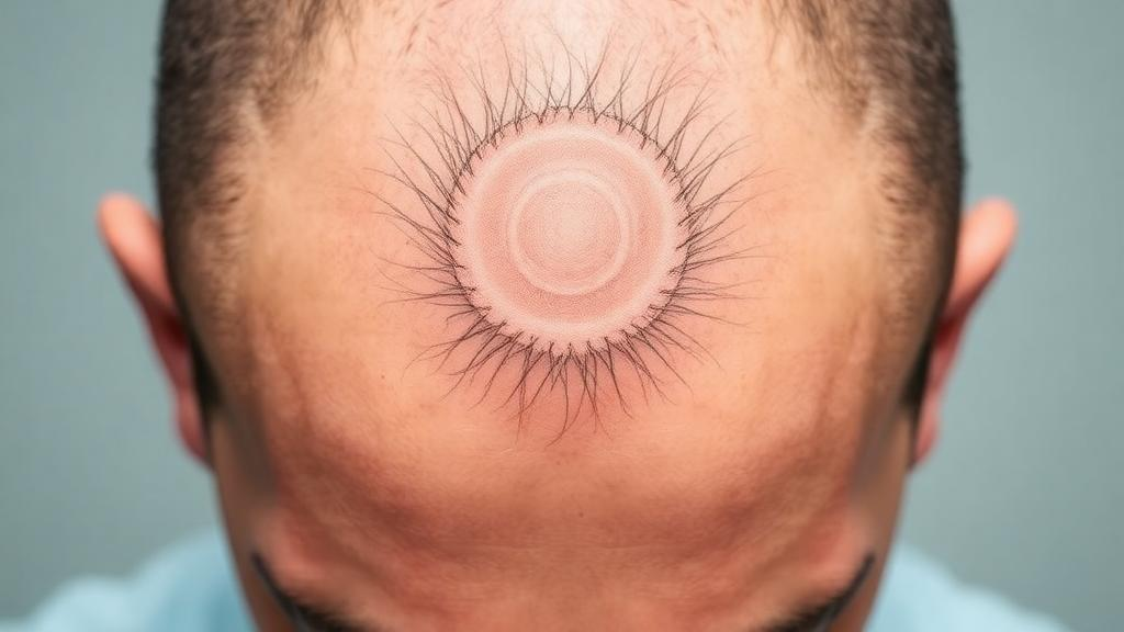
A single case from Japan is drawing attention for what it reveals about a disfiguring scalp condition that stubbornly resisted two of modern dermatology’s most powerful drugs. Dermatologists at Kindai University Hospital in Osaka describe a man in his 40s whose occipital scalp had been ravaged for eight years by dissecting cellulitis of the scalp, an inflammatory disorder closely related to hidradenitis suppurativa. Despite a full year of adalimumab followed by six months of bimekizumab, two biologic agents that routinely transform the lives of patients with hidradenitis suppurativa, his lesions showed no meaningful improvement. The reason, the clinicians argue, may have been hiding in plain sight beneath the surface of his skin: masses of hair shafts trapped inside the diseased tissue itself.
Dissecting cellulitis of the scalp belongs to a family of conditions known as the follicular occlusion disorders, a group that also includes hidradenitis suppurativa, acne conglobata and pilonidal disease. All of them share a fundamental defect in which hair follicles become plugged and rupture, unleashing chronic inflammation deep within the skin. On the scalp, this process produces recurrent painful nodules, abscesses and interconnected tunnels called sinus tracts that weep pus and eventually heal with dense scarring. Over time, the disease can destroy hair follicles wholesale, producing patches of permanent scarring alopecia. While biologic drugs that block inflammatory pathways such as tumor necrosis factor and interleukin-17 have become mainstays of hidradenitis suppurativa therapy, their track record in the scalp-specific variant remains poorly defined, and treatment algorithms for dissecting cellulitis rely largely on small series and expert opinion rather than rigorous trials.
The patient’s story began with nodular lesions on the back of his head that slowly enlarged over eight years, becoming painful, discharging pus and steadily eroding his hair. He received his initial diagnosis of hidradenitis suppurativa at an outside clinic, where the biologic trials were undertaken. By the time he reached the Osaka team, the occipital scalp carried an extensive plaque of scarring alopecia measuring roughly 13 by 13 centimeters, studded with tufted hair, a striking morphological pattern in which multiple hair shafts emerge from a single dilated follicular opening like strands gathered through one buttonhole. The area was red, firm and indurated, with purulent material draining from sinus tracts that riddled the affected skin.
The clinicians first attempted incision and drainage followed by deroofing, a surgical technique in which the roofs of inflamed tunnels are removed to allow healing from the base. The procedure produced only limited improvement, but it exposed something unexpected: elongated hair shafts extending within the subcutaneous tissue, protruding from the wound. This finding prompted the team to proceed with wide excision of the entire affected region, and the cut surface of the removed specimen told the story plainly. The fibrotic tissue beneath the scalp was markedly thickened, and the sinus tracts within it were packed with numerous retained hair shafts, a physical record of years of follicular destruction and failed hair extrusion.
Histopathological examination of the excised tissue confirmed the clinical impression. Under the microscope, the follicular structures had been destroyed, inflammatory cells infiltrated the dermis, and bands of fibrosis fused adjacent follicles together, a picture consistent with scarring alopecia. The tufted hair visible on the surface is now understood as a downstream consequence of this architecture: when the upper portions of follicles are destroyed and fibrosis welds neighboring openings together, several surviving hair shafts exit the skin through a single shared orifice. The postoperative course was uneventful, and the patient experienced no recurrence after surgery.
The most consequential observation, however, is the one the authors highlight as unusual: the accumulation of hair shafts within the sinus tracts themselves. Normally, hair shafts that lose their follicles are shed from the skin surface or expelled through inflammatory channels. In this patient, follicular destruction and surrounding fibrosis appear to have impaired that extrusion process, leaving shafts stranded within the subcutaneous tissue. Retained hair is a known foreign body and a reservoir for bacterial colonization, and its persistence inside closed tracts offers a plausible mechanical explanation for why the disease kept flaring regardless of how effectively the biologics suppressed inflammatory signaling.
That interpretation carries a broader lesson about the limits of immunology-directed therapy. Biologic agents excel at controlling the immune-driven components of inflammatory skin disease, but they cannot dissolve scar tissue, reopen obliterated follicles or remove physical debris lodged within fistulas. The authors suggest that in fibrosis-dominant lesions such as established scarring and mature sinus tracts, the efficacy of biologics may be inherently limited. The presence of scarring alopecia, tufted hair and intralesional hair accumulation in this patient indicated that the disease had already progressed to a structurally remodeled, fibrosis-dominant stage by the time biologic treatment began, a stage at which anti-inflammatory drugs alone were unlikely to succeed.
The case therefore offers clinicians a practical diagnostic clue as much as a therapeutic one. When a patient with dissecting cellulitis presents with scarring alopecia and tufted hair, the authors advise that fibrosis-dominant disease with underlying structural abnormalities, including subcutaneous hair retention, should be suspected. In such presentations, the response to biologic therapy may be limited, and early consideration of surgical excision is warranted rather than prolonged cycles of escalating medical therapy. The reasoning echoes a principle familiar across inflammatory dermatology: timing matters, and interventions aimed at inflammation deliver their greatest benefit before fibrosis has permanently rewritten the tissue architecture.
For the wider field, the report adds a concrete morphological marker to the still-fragmentary picture of dissecting cellulitis, a rare condition for which no randomized controlled trials have established a standard of care. Systematic reviews of published treatments have catalogued antibiotics, retinoids, corticosteroids, biologics and assorted surgical approaches, but the evidence base remains thin. Case reports like this one, in which the excised tissue can be examined in full cross-section, provide the granular anatomical detail that larger studies often lack, and they help generate the hypotheses that future trials must test, such as whether early surgical clearance of fibrosis-dominant disease outperforms extended biologic courses in patients with advanced scarring.
The report also underscores the value of careful surgical inspection. Had the team not proceeded to wide excision, the hair-filled sinus tracts would have remained invisible, and the failure of biologic therapy might have been attributed to drug resistance or misdiagnosis rather than to the physical anatomy of the disease. For patients with dissecting cellulitis whose lesions fail to respond to state-of-the-art biologics, the message from Osaka is that the answer may lie not in another injection but in what the scalpel reveals: trapped hair, dense fibrosis and tunnels that no immune-modulating drug can unmake.
Subject of Research: A case report of dissecting cellulitis of the scalp with hair retention in sinus tracts and limited response to biologic therapy
Article Title: Dissecting cellulitis of the occipital scalp with hair retention within sinus tracts
Article References: Dissecting cellulitis of the occipital scalp with hair retention within sinus tracts. (n.d.). https://doi.org/10.3389/jcia.2026.17140
Image Credits: AI Generated
Keywords: dissecting cellulitis, hidradenitis suppurativa, sinus tracts, tufted hair, scarring alopecia, follicular occlusion, biologic therapy, adalimumab, bimekizumab, surgical excision, dermatology, case report
Cite Scienmag News
Ophelia Keating. (October 16, 2026). Trapped Hairs Inside Sinus Tracts May Explain Why a Severe Scalp Disease Resisted Biologics. Scienmag. https://scienmag.com/trapped-hairs-inside-sinus-tracts-may-explain-why-a-severe-scalp-disease-resisted-biologics/
Ophelia Keating. "Trapped Hairs Inside Sinus Tracts May Explain Why a Severe Scalp Disease Resisted Biologics." Scienmag, 16 October 2026, https://scienmag.com/trapped-hairs-inside-sinus-tracts-may-explain-why-a-severe-scalp-disease-resisted-biologics/. Accessed 16 October 2026.
Ophelia Keating. "Trapped Hairs Inside Sinus Tracts May Explain Why a Severe Scalp Disease Resisted Biologics." Scienmag. October 16, 2026. https://scienmag.com/trapped-hairs-inside-sinus-tracts-may-explain-why-a-severe-scalp-disease-resisted-biologics/


