


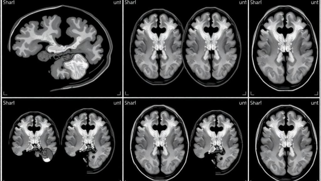
Every year, millions of newborns around the world experience perinatal asphyxia, a dangerous drop in oxygen supply during the process of birth. Even when babies survive the acute crisis, the injury left behind in their developing brains can echo across a lifetime, producing anything from subtle developmental delays to cerebral palsy. Now a team of researchers at the Children’s Hospital of Soochow University in Suzhou, China, has identified a strikingly simple marker visible on ordinary magnetic resonance imaging that may help clinicians judge how badly an asphyxiated newborn’s brain has been damaged and even forecast whether that child will struggle with sleep years down the road. The marker is the enlarged perivascular space, a fluid-filled channel that surrounds blood vessels deep in the brain, and its appearance in large numbers on neonatal scans is emerging as a window into a biological system that scientists have only recently come to appreciate: the glymphatic network that washes waste out of the brain.
Perivascular spaces are, in healthy brains, almost invisible. They are the cuffs of cerebrospinal fluid that accompany penetrating arteries and veins into the brain’s substance, and in a typical newborn the amount of fluid within them is too small to register on clinical imaging. When these spaces balloon and multiply, however, they show up on T2-weighted magnetic resonance images as bright, linear or round dots, and on FLAIR sequences as dark counterparts of the same shapes. In adults, a heavy burden of enlarged perivascular spaces has been linked to cerebral small vessel disease, cognitive decline, and poor glymphatic clearance of toxic proteins. Until now, almost nothing was known about what these structures mean in the earliest days of life, particularly in babies whose brains have just endured the twin assaults of oxygen deprivation and the inflammatory firestorm that follows it.
The new study, published in the World Journal of Pediatrics, enrolled 319 newborns admitted to the hospital’s neonatology department between January 2020 and September 2023, all transferred within a week of birth. Using strict diagnostic criteria based on Apgar scores and umbilical artery blood gas analysis, the researchers divided the infants into a mild asphyxia group of 229 babies and a severe group of 90. Each child underwent brain magnetic resonance imaging on a 3.0 Tesla scanner, along with amplitude-integrated electroencephalography to capture the electrical rhythms of the injured cortex, and a battery of laboratory tests ranging from inflammatory markers to coagulation profiles. Two experienced radiologists, blinded to all clinical information, counted the enlarged perivascular spaces in the basal ganglia across four to six axial sections, producing a quantitative burden score for every infant.
The results were unambiguous. In the severe asphyxia group, lactate levels and C-reactive protein concentrations were significantly elevated compared with the mild group, and the number of enlarged perivascular spaces was also markedly higher. Across the whole cohort, the count of these spaces correlated positively with diastolic blood pressure and lactate, and negatively with Apgar scores at one, five, and ten minutes. More importantly, when the researchers built multivariable linear regression models to disentangle independent predictors, the number of enlarged perivascular spaces remained significantly associated with the severity of brain injury as graded on a simplified magnetic resonance score, with a P value of 0.0001 across all infants, 0.0248 in the mild group, and 0.0114 in the severe group. This held true even after accounting for gestational age, blood pressure, base excess, Apgar scores, and coagulation measures such as thrombin time and the international normalized ratio.
Intriguingly, the association was selective. The perivascular space count predicted the structural magnetic resonance score but not the amplitude-integrated electroencephalography grade, a measure of acute cortical electrical function. The authors interpret this dissociation as evidence that enlarged perivascular spaces capture structural, glymphatic damage rather than transient functional suppression. Electroencephalographic activity can be temporarily blunted by metabolic derangement and then recover, whereas the magnetic resonance image visualizes established injury to brain parenchyma. A marker of perivascular structural integrity, the researchers argue, aligns more naturally with the latter. This distinction matters clinically, because it suggests the marker adds a dimension of information that neither routine electrophysiology nor blood-based biomarkers of inflammation and neuronal injury can currently provide.
The mechanistic story behind the finding centers on the glymphatic system, the recently characterized network through which cerebrospinal fluid pulses along perivascular channels to flush metabolic waste from the brain, driven in part by the water channel protein aquaporin 4, which is polarized to the endfeet of astrocytes. Asphyxia triggers oxidative stress and a cytokine storm that disrupt this polarization, causing perivascular spaces to enlarge and glymphatic clearance to falter. The consequence may be self-reinforcing: as clearance fails, neurotoxic metabolites accumulate, further amplifying inflammation and astrocytic injury in a vicious cycle of progressive damage. The authors are careful to note that their observational design cannot establish causality; the enlarged spaces may actively contribute to injury or may simply be an epiphenomenon of the same upstream pathology. Either way, their count offers a non-invasive readout of a process that has been nearly impossible to monitor in living newborns.
Perhaps the most provocative result concerns sleep. Because mild asphyxial injury can damage the pineal gland, the source of melatonin, sleep disturbance has long been recognized as a sequela of perinatal asphyxia. The Suzhou team followed 258 of the 319 infants, using monthly parental questionnaires over six months, adapted from the Brief Infant Sleep Questionnaire and the Paediatric Sleep Questionnaire, to score seven sleep problems including prolonged sleep latency, insufficient sleep, irregular schedules, snoring, breathing difficulties, and frequent night wakings. After excluding age as a confounder by stratifying children into three age bands, the researchers found no relationship between perivascular space count and sleep in the full cohort or in the mild group. But in the severe asphyxia group, the correlation was significant: a higher number of enlarged perivascular spaces predicted more severe sleep disturbance, with a Spearman coefficient of 0.3365 and a P value of 0.0061.
That severity-specific pattern, the authors suggest, points to a mechanism beyond direct pineal injury. In severely asphyxiated infants, glymphatic damage may impede the clearance of toxic proteins, perpetuating brain injury and indirectly degrading sleep architecture, whereas in mild cases the perivascular spaces tend to shrink over time as the infant brain repairs itself, explaining the absence of a sleep correlation. The correlation is moderate rather than overwhelming, and the researchers acknowledge that infant sleep is multifactorial and cannot be reduced to a single biomarker. Still, the finding hints that a routine scan performed in the first week of life could flag babies at risk of sleep problems before parents notice anything wrong, opening a window for early behavioral and medical interventions during a period when sleep is critical to brain development.
The study has limitations that the authors confront directly. Ethical constraints precluded a healthy control group, though published data confirm that enlarged perivascular spaces are rare in healthy neonates, and the single-center design, moderate sample size, and reliance on parent-reported sleep questionnaires all warrant caution. The team calls for multicenter prospective cohorts incorporating diffusion tensor imaging along the perivascular space, an advanced technique that quantifies glymphatic flow directly, along with objective sleep monitoring and longitudinal follow-up. If those studies confirm the present findings, the humble perivascular space, long dismissed as an incidental dot on adult brain scans, could become a standard element of neonatal risk stratification, helping clinicians decide which asphyxiated newborns need the most aggressive neuroprotective care and which families should be counseled and monitored for the sleep disorders that may otherwise surface silently in the years after a difficult birth.
Subject of Research: Enlarged perivascular spaces as imaging biomarkers of perinatal asphyxia-induced brain injury and later sleep quality
Article Title: Correlation between perinatal asphyxia-induced brain injury, number of enlarged perivascular spaces, and sleep quality in later life
Article References: Xue, X.-Y., Sun, L., Yu, M., Li, M.-X., Tian, Q.-Y., Ren, J., Miao, P., Xu, L.-X., Feng, C.-X., Li, M., Gong, M., Wang, L.-Z., Huo, H.-L., Huang, J., Guo, W.-L., Ju, R., Pan, T., Feng, X., Zhou, W.-H., … Ding, X. (2026). Correlation between perinatal asphyxia-induced brain injury, number of enlarged perivascular spaces, and sleep quality in later life. World Journal of Pediatrics, 22(9), 1055-1068. https://doi.org/10.1007/s12519-026-01093-8
Image Credits: AI Generated
DOI: 10.1007/s12519-026-01093-8
Keywords: perinatal asphyxia, enlarged perivascular spaces, glymphatic system, neonatal brain injury, MRI biomarker, sleep quality, hypoxic-ischemic encephalopathy, newborns, aquaporin 4, neonatology, amplitude-integrated EEG, World Journal of Pediatrics
Cite Scienmag News
Harold Sullivan. (September 30, 2026). Tiny Brain Spaces on Newborn Scans May Predict Sleep Trouble Years Later. Scienmag. https://scienmag.com/tiny-brain-spaces-on-newborn-scans-may-predict-sleep-trouble-years-later/
Harold Sullivan. "Tiny Brain Spaces on Newborn Scans May Predict Sleep Trouble Years Later." Scienmag, 30 September 2026, https://scienmag.com/tiny-brain-spaces-on-newborn-scans-may-predict-sleep-trouble-years-later/. Accessed 16 October 2026.
Harold Sullivan. "Tiny Brain Spaces on Newborn Scans May Predict Sleep Trouble Years Later." Scienmag. September 30, 2026. https://scienmag.com/tiny-brain-spaces-on-newborn-scans-may-predict-sleep-trouble-years-later/


