In the intricate world of cellular biology, the mechanics of wound healing have long captivated scientists, weaving together threads of biochemical signaling and cellular movement. A groundbreaking study from the Tata Institute of Fundamental Research (TIFR), Hyderabad, has unveiled a previously unseen cellular protagonist that directs how epithelial cells respond to the geometry of wound edges. This research spotlights the endoplasmic reticulum (ER)—the largest intracellular organelle—not merely as a biosynthetic powerhouse but as a dynamic mechanosensor that reads the curvature of a wound gap and orchestrates the mode of epithelial migration during tissue repair.
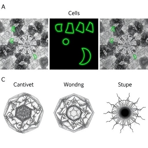
When skin sustains an injury, the epithelial cells bordering the wound engage in a highly coordinated effort to close the gap. Although this phenomenon is well-known, what remained elusive until now was how cells interpret the subtle, microscopic curvatures at the wound boundary to adopt distinct migration strategies. Remarkably, the pioneering research led by graduate student Simran Rawal within Tamal Das’s laboratory demonstrates that the shape of the wound edge — whether convex or concave — dramatically reshapes the architecture of the ER within epithelial cells. Such morphological adaptations result in fundamentally different cellular behaviors that influence the entire wound closure process.
The ER’s structural shifts are profound. At convex wound edges, the ER transitions into a tubular, network-like form, sprawling towards the cell periphery near the gap. Conversely, near concave boundaries, the ER adopts a flattened, sheet-like configuration. This reorganization is not a mere structural curiosity; it actively dictates whether cells will extend membrane protrusions and crawl into the wound space or contract collectively, employing a purse-string mechanism to tighten the wound margins.
Delving deeper into the cellular machinery, Rawal and colleagues observed that these ER morphological transitions depend critically on the dynamics and interplay of the cytoskeleton—specifically, microtubules and actin filaments. At concave interfaces, a balance of both cytoskeletal components modulates the flattened ER sheet formation. In contrast, convex edges rely predominantly on microtubules to stabilize the ER’s tubular morphology. By experimentally perturbing the ER shape—forcing cells at concave edges to harbor tubular instead of sheet-like ER—the team convincingly demonstrated a switch in migratory behavior from contraction-based closure to crawling, underscoring the causal role of ER topology in cell motility decisions.
The study also illuminated the mechanical underpinnings governing these phenomena through a collaboration with researchers at the University of Birmingham. Here, mathematician Pradeep Keshavanarayana developed computational models calculating strain energy within cells confronted with diverse gap curvatures. Their results suggest that ER reorganization minimizes intracellular strain energy, optimizing mechanical efficiency during migration. In essence, the ER doesn’t just respond passively but adapts in a manner that fine-tunes cellular biomechanics requisite for effective wound sealing.
Perhaps the most remarkable insight is the ER’s emerging role as a mechanotransducer that bridges mechanical inputs and biochemical signaling pathways. Spanning the cell from the nuclear envelope to the periphery, the ER’s structural plasticity enables it to distribute mechanical cues internally, potentially triggering cascades that influence gene expression, cytoskeletal dynamics, and membrane trafficking. Such integrative functionalities position the ER at the nexus of physical and molecular cell biology during tissue regeneration.
This discovery challenges traditional dogma that has largely confined organelle function to biochemical roles such as calcium handling and protein synthesis. Instead, it extends the functional repertoire of the ER to include sensing and responding to geometric cues at the tissue scale, emphasizing the importance of cellular architecture in developmental biology and regenerative medicine. The research pivots the scientific gaze towards a more holistic perspective, where the physical microenvironment and organelle dynamics synergize to direct collective cell behavior.
Beyond wound healing, these findings open uncharted avenues for exploring how intracellular organelles might govern tissue formation and repair in diverse biological contexts. Could the ER’s curvature-sensitive responses influence morphogenesis during embryonic development? Might similar mechanosensory roles be at play in other organelles or in pathologies involving aberrant tissue remodeling? The tantalizing prospect emerges that intracellular organelles have underappreciated roles as spatial and mechanical sensors influencing multicellular organization.
The key to this intricate cellular dance lies in understanding how mechanical strain induced by changes in extracellular geometry is propagated and decoded internally. The ER, acting as a continuous membrane system tethered to the cytoskeleton, is uniquely positioned to sustain and transmit these mechanical signals over long intracellular distances. This capacity likely enables epithelial sheets to coordinate collective responses, ensuring robust wound closure despite the variable and complex shapes that natural wounds assume.
Simran Rawal’s meticulous live-cell imaging and structural analyses were pivotal in capturing the ER’s dynamic remodeling in real time. Complemented by cytoskeletal perturbation experiments and mathematical modeling, the study exemplifies the power of multidisciplinary approaches to dissect complex biological processes. Such synergy between experimental and theoretical frameworks provides compelling mechanistic explanations that transcend purely descriptive observations.
As this thread of research unfolds, it beckons a reevaluation of tissue engineering strategies and wound management therapies. Manipulating intracellular organelle morphology or modulating mechanical feedback pathways may emerge as innovative avenues to enhance tissue repair. Furthermore, this new understanding invigorates the exploration of intracellular mechanics as essential determinants of cellular fate decisions, a frontier with profound implications for regenerative medicine and cancer biology alike.
In sum, this landmark study redefines the endoplasmic reticulum not just as a cellular organelle but as a pivotal sensor and mediator that deciphers wound edge geometry to steer epithelial migration modes. The meticulous uncovering of this mechanism profoundly enriches our knowledge of how cells transmute physical landscapes into biochemical instructions, orchestrating the harmonious choreography vital for tissue restoration. As researchers worldwide digest these insights, the ER’s curvature-sensitive capabilities may emerge as a central theme in the nexus between cell biology, biophysics, and regenerative therapeutics.
Subject of Research: Cell biology and mechanotransduction in epithelial wound healing
Article Title: Edge curvature drives endoplasmic reticulum reorganization and dictates epithelial migration mode
Web References:
- Tata Institute of Fundamental Research, Hyderabad
- University of Birmingham
- Nature Cell Biology Article
- DOI: 10.1038/s41467-024-53207-3
References: Rawal S., Das T., Keshavanarayana P., et al. “Edge curvature drives endoplasmic reticulum reorganization and dictates epithelial migration mode.” Nature Cell Biology, 2024.
Image Credits: Simran Rawal, Tata Institute of Fundamental Research, Hyderabad, India
Keywords: Endoplasmic reticulum, epithelial migration, wound healing, mechanotransduction, cell morphology, cytoskeleton dynamics, curvature sensing, tissue repair, microtubules, actin filaments, intracellular strain, cell motility
