


Bone and Soft Tissue Combined: New Imaging Method Builds 3D Shape Models of the Foot and Ankle
A new method combines weight-bearing CT and MRI to build reliable three-dimensional statistical shape models of foot and ankle bones, ...
A new method combines weight-bearing CT and MRI to build reliable three-dimensional statistical shape models of foot and ankle bones, ...
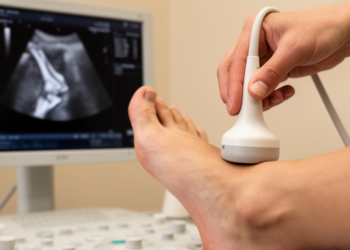
A cadaveric validation study found that ultrasonography located the peroneus brevis tendon insertion at the fifth metatarsal base with a ...
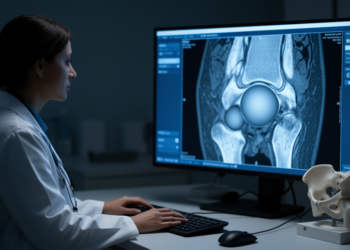
Three-dimensional CT analysis of 22 FAI patients found no significant pre- to postoperative shift of the femoral head center after ...

A cross-sectional ultrasound and shear-wave elastography study of 45 racket-sport players and 45 matched controls found significantly thicker and stiffer ...
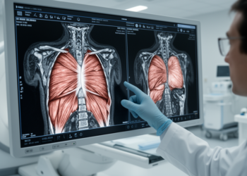
Researchers have used high-resolution MRI and manual segmentation to measure, for the first time, the fascicle length, volume, orientation, and ...
A new review in Sports Medicine - Open shows that dynamic ultrasound, Doppler imaging, and elastography can reveal the structure, ...
A new Pediatric Radiology case report describes how ultrasound revealed a Salter-Harris type I fracture-dislocation of the distal phalangeal epiphysis ...


Embark on a thrilling journey of discovery with Scienmag.com—your ultimate source for cutting-edge breakthroughs. Immerse yourself in a world where curiosity knows no limits and tomorrow’s possibilities become today’s reality!