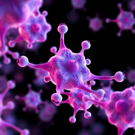
In a groundbreaking advancement for cellular biology and personalized medicine, researchers have unveiled the HCS-3DX platform, a revolutionary high-content screening system designed specifically for the automated and AI-driven analysis of three-dimensional multicellular structures such as spheroids and organoids. This state-of-the-art technology promises to dramatically accelerate drug discovery, mechanobiology research, and regenerative medicine by enabling researchers to perform high-precision, single-cell resolution imaging within complex 3D tissue models at an unprecedented throughput.
The HCS-3DX system integrates artificial intelligence algorithms into the image processing and segmentation pipeline, allowing it to identify and analyze individual cells throughout intricate 3D cell cultures. This is a significant leap forward compared to traditional two-dimensional assays or less sophisticated 3D imaging methods which often struggle with cell overlap, image artifacts, and lower resolution. By providing fully automated workflows from sample preparation to data extraction, the system substantially reduces manual intervention and operator bias, enhancing the reproducibility and scalability of experiments.
At the heart of the platform lies an innovative modular architecture that permits seamless adaptation to a wide variety of experimental setups and throughput demands. Researchers can configure the system to accommodate different sizes of spheroids and organoids, diverse staining protocols, and multiple imaging modalities including fluorescence and brightfield. This flexibility is crucial for broad applicability across diverse fields, such as oncology, developmental biology, and toxicology, where cellular heterogeneity and microenvironmental context play critical roles.
Ákos Diósdi, the principal architect of the platform, emphasized the importance of creating a unified solution that amalgamates the strengths of existing 3D culture analysis tools while overcoming their limitations. The AI-powered framework not only expedites image acquisition and processing but also enhances accuracy by robustly discerning cellular boundaries, subcellular features, and phenotypic biomarkers within dense tissue-like structures. This capability ensures that large data volumes, often extending to thousands of cells per structure, can be analyzed swiftly and with meticulous granularity.
One of the chronic bottlenecks in personalized medicine has been the limited throughput and scalability of functional cell-based assays, restricting the ability to screen therapeutic compounds rapidly across patient-derived models. According to Dr. Péter Horváth, director at the HUN-REN Biological Research Centre, the HCS-3DX platform effectively overcomes these constraints. Its accelerated screening capability allows clinicians and scientists to generate highly accurate, individualized drug response profiles within clinically relevant timeframes, potentially guiding more effective patient-specific treatment regimens.
The platform’s impact is already being demonstrated in translational research collaborations, notably with the Heidelberg Children’s Hospital. There, miniature tumor models derived from pediatric brain cancer patients are cultured as organoids and subjected to high-content screening on the HCS-3DX system. This approach enables the identification of the most efficacious therapeutic candidates by evaluating drug responses on a single-cell basis, shedding light on intratumoral heterogeneity and resistance mechanisms that would otherwise remain concealed in bulk assays.
Technical innovations incorporated in the system include advanced 3D imaging optics combined with optimized AI segmentation algorithms that facilitate the extraction of multidimensional morphological and molecular features. This high-dimensional data enables the detailed characterization of cellular phenotypes, proliferation, apoptosis, and spatial cellular interactions, all within the physiologically relevant 3D contexts that better mimic in vivo conditions compared to monolayer cultures.
Furthermore, the automated workflow encompasses intelligent sample selection processes, ensuring that only high-quality spheroids meeting predefined morphological criteria enter the imaging pipeline. This quality control measure prevents wasted resources on analyzing suboptimal samples and enhances the statistical robustness of experimental outcomes. Together, these capabilities position the HCS-3DX as a transformative tool for both fundamental research and pharmaceutical development workflows.
As automated 3D cell culture analysis becomes increasingly essential for understanding tissue complexity and disease biology, the emergence of platforms like HCS-3DX marks a vital turning point. It empowers researchers with rapid, scalable, and precise data acquisition that can uncover previously inaccessible mechanistic insights into multicellular dynamics, morphogenesis, and drug action at single-cell resolution within intact biological models.
Given the pressing demand for improved preclinical models and precision therapeutics, HCS-3DX’s combination of AI-driven segmentation, modular adaptability, and clinically translatable screening holds significant promise for shaping the future landscape of biomedical research and personalized healthcare. The platform is poised to facilitate novel discoveries that translate directly to better patient outcomes, reflecting a new era of intégrated, high-throughput 3D biological analysis.
By streamlining the convergence of cutting-edge microscopy, computational intelligence, and tissue engineering, the HCS-3DX system exemplifies the potential of technology-driven innovation to surmount longstanding scientific challenges. As researchers worldwide adopt this solution, the increased throughput and fidelity of 3D high-content screening may well accelerate the development of targeted therapies for complex diseases, ultimately bridging the gap between laboratory bench and clinical bedside more effectively than ever before.
Subject of Research: Cells
Article Title: HCS-3DX, a next-generation AI-driven automated 3D-oid high-content screening system
News Publication Date: 7-Oct-2025
Web References: http://dx.doi.org/10.1038/s41467-025-63955-5
References: 10.1038/s41467-025-63955-5
Image Credits: Akos Diosdi
Keywords: AI-driven imaging, high-content screening, 3D cell cultures, spheroids, organoids, automated microscopy, personalized medicine, drug screening, cellular heterogeneity, regenerative medicine, image segmentation, biomedical innovation


