


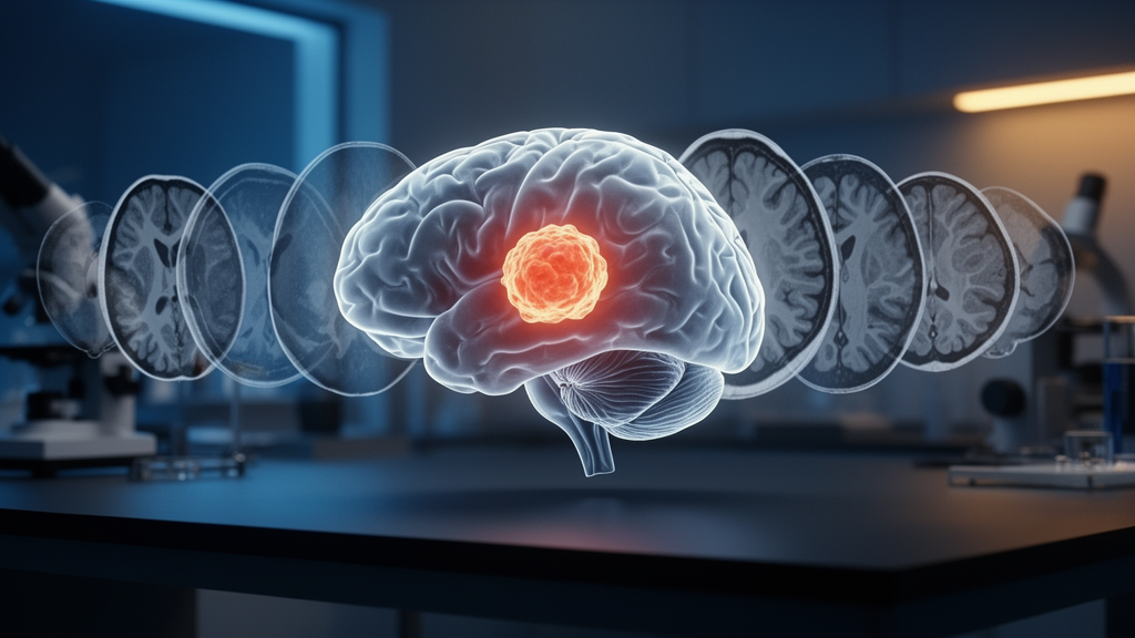
Deep learning has become one of the most powerful tools in modern radiology, but the performance of any neural network ultimately depends on what it is fed. A new study published in BMC Medical Imaging suggests that a relatively simple change to the input data, rather than a radical redesign of the network itself, may be enough to sharpen the automated delineation of brain tumors. Amir Khorasani of Isfahan University of Medical Sciences in Iran reconstructed synthetic relative T1/T2 (rT1/T2) maps from routine magnetic resonance imaging sequences and used them as the sole input channel for U-Net models tasked with segmenting glioma sub-regions, with results that consistently edged out conventional T1-weighted and T2-weighted images.
Gliomas are the most common primary brain tumors in adults, and their precise segmentation on MRI is far more than an academic exercise. Radiologists and neurosurgeons rely on the boundaries of three distinct sub-regions, the peritumoral edema that swells around the tumor mass, the necrotic core where tissue has died, and the active tumor where proliferating cells drive disease progression. Treatment monitoring and surgical planning both hinge on how accurately these compartments are outlined. The gold standard in machine learning research, the BraTS challenge dataset, has fueled years of progress in automated segmentation, yet the field continues to search for input representations that expose tumor biology more clearly to the algorithms doing the work.
The idea behind rT1/T2 maps is rooted in quantitative MRI physics. T1 and T2 are intrinsic relaxation properties of tissue, describing how quickly hydrogen nuclei recover their magnetization after excitation and how rapidly the signal decays, respectively. Different tissues relax at different rates, which is why a glioma looks different from healthy white matter on a scanner. Rather than feeding the network the raw weighted images, whose intensities depend on scanner settings and acquisition protocols, the study reconstructed a pixel-wise ratio of the two relaxation properties. This relative map is a synthetic modality, computed from preprocessed T1-weighted and T2-weighted sequences, and it condenses complementary information from both acquisitions into a single normalized channel.
To test whether this synthetic input actually helps, Khorasani trained backbone U-Nets, the workhorse architecture of medical image segmentation, on 381 cases drawn from the BraTS 2023 dataset. The U-Net design pairs an encoder that compresses spatial information with a decoder that reconstructs fine-grained masks, and its performance can be tuned by swapping the backbone network that performs the feature extraction. Six well-known convolutional backbones were put to the test: ResNet34, ResNet152, InceptionV3, VGG16, DenseNet121, and MobileNet. Each model was trained in a single-channel configuration, meaning the network saw only the rT1/T2 map rather than the multi-sequence stacks common in clinical pipelines, and evaluation used 5-fold cross-validation with patient-wise train-test splitting to ensure that no patient’s scans appeared in both training and testing data.
Performance was measured with three complementary metrics. The Dice Similarity Coefficient quantifies the overlap between the predicted mask and the ground truth, ranging from zero for no agreement to one for perfect overlap. Intersection over Union penalizes both false positives and false negatives in a single ratio. The 95th percentile Hausdorff Distance, abbreviated HD95, captures how far the predicted boundary strays from the true boundary in millimeters, focusing on the worst 5 percent of surface points to avoid being fooled by a few outlier pixels. Across the peritumoral edema, necrotic core, and active tumor regions, these metrics together paint a picture of both volumetric accuracy and geometric precision.
The results favored the synthetic maps. A ResNet34 backbone U-Net trained on rT1/T2 maps achieved the best performance for edema segmentation, with a Dice score of 0.901, an HD95 of 1.57 millimeters, and a correlation coefficient of 0.997. The same architecture led on the necrotic core, reaching a Dice of 0.816, an HD95 of 2.43 millimeters, and a correlation of 0.994. For the hardest target, the actively proliferating tumor, an InceptionV3 backbone U-Net took the lead with a Dice of 0.837, an HD95 of 1.66 millimeters, and a correlation of 0.975. These numbers indicate that the networks were not merely capturing coarse tumor extent but resolving biologically meaningful internal structure.
To rank the many combinations of backbone and input modality objectively, the study applied TOPSIS, the Technique for Order of Preference by Similarity to Ideal Solution, a multi-criteria decision analysis method that scores each candidate by its geometric distance to an ideal and an anti-ideal solution. In this ranking, rT1/T2 maps placed first overall with a closeness coefficient of 0.794, and InceptionV3 emerged as the top-ranked backbone with a closeness coefficient of 0.938. The analysis provides a systematic answer to a question that single-metric comparisons often leave ambiguous, namely which pairing of architecture and input delivers the best balance across all three tumor sub-regions and all evaluation metrics simultaneously.
The author is careful to frame the finding with appropriate scientific caution. Within this single-channel experimental setting, the rT1/T2 maps showed what the study describes as a modest but consistent average advantage over conventional T1-weighted and T2-weighted sequences across the backbone architectures tested. Crucially, the study leaves open whether that advantage stems from the ratio transformation itself, which may highlight tissue contrast in a way the raw sequences cannot, or simply from the fact that the synthetic map draws on information contained in both parent sequences. The appropriate control, a multi-channel input combining T1 and T2 side by side, was not tested here and is flagged as a priority for future work.
The implications for clinical practice are nonetheless intriguing. Single-channel inputs reduce memory demands and computational cost, which matters for deploying segmentation models in hospitals with limited hardware or for integrating them into real-time surgical navigation. A synthetic modality derived from sequences that every glioma patient already receives adds no scanning burden, no contrast agent, and no acquisition time. If the advantage holds up against multi-channel baselines and external datasets, ratio-based inputs could become a standard preprocessing step rather than a research curiosity.
The work also fits into a broader trend in medical AI, where the spotlight is shifting from ever-larger networks toward smarter representations of the data themselves. Quantitative and semi-quantitative maps, including relaxation ratios, diffusion-derived metrics, and perfusion parameters, encode tissue physiology more directly than conventional weighted images. As segmentation models are increasingly asked to support treatment monitoring and surgical planning for glioblastoma and other gliomas, the choice of input modality may prove as consequential as the choice of architecture. This study, supported by Isfahan University of Medical Sciences and approved under the Declaration of Helsinki, offers a measured but compelling data point in that evolving conversation, and its open-access publication ensures that other teams can immediately test the idea on their own data.
Subject of Research: Synthetic rT1/T2 maps as a novel input modality for backbone U-Net glioma sub-region segmentation
Article Title: Synthetic rT1/T2 maps as a novel input modality for backbone U-Net glioma sub-region segmentation
Article References: Khorasani, A. (2026). Synthetic rT1/T2 maps as a novel input modality for backbone U-Net glioma sub-region segmentation. BMC Medical Imaging. https://doi.org/10.1186/s12880-026-02782-6
Image Credits: AI Generated
DOI: 10.1186/s12880-026-02782-6
Keywords: glioma, brain tumor segmentation, deep learning, U-Net, MRI, rT1/T2 maps, BraTS dataset, ResNet, InceptionV3, TOPSIS, medical imaging, artificial intelligence
Cite Scienmag News
Cassandra Pierce. (October 18, 2026). Synthetic rT1/T2 Maps Give U-Net Networks a Sharper View of Brain Tumors. Scienmag. https://scienmag.com/synthetic-rt1-t2-maps-give-u-net-networks-a-sharper-view-of-brain-tumors/
Cassandra Pierce. "Synthetic rT1/T2 Maps Give U-Net Networks a Sharper View of Brain Tumors." Scienmag, 18 October 2026, https://scienmag.com/synthetic-rt1-t2-maps-give-u-net-networks-a-sharper-view-of-brain-tumors/. Accessed 18 October 2026.
Cassandra Pierce. "Synthetic rT1/T2 Maps Give U-Net Networks a Sharper View of Brain Tumors." Scienmag. October 18, 2026. https://scienmag.com/synthetic-rt1-t2-maps-give-u-net-networks-a-sharper-view-of-brain-tumors/


