Unveiling Hidden Signals: Lung Cancer Screenings Reveal Clues to Other Cancers
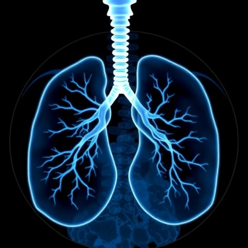
In the evolving landscape of medical diagnostics, the use of computerized tomography (CT) scans to detect lung cancer has become a cornerstone for screening high-risk populations, particularly among chronic smokers. However, recent research spearheaded by the Brown University School of Public Health has illuminated a compelling secondary benefit of this technology: the incidental detection of abnormalities beyond the pulmonary system that may signal other, previously undiagnosed malignancies. This groundbreaking study leverages data from the National Lung Screening Trial (NLST), a monumental federal clinical trial involving over 26,000 participants, and scrutinizes the implications of findings unrelated to lung cancer in these scans.
Traditionally, CT scanning was embraced for its capacity to visualize lung nodules with exceptional clarity, facilitating early intervention in lung cancer cases and substantially improving patient outcomes. What has become increasingly apparent, and is now meticulously explored by Brown researchers, is the clinical significance of incidental findings—abnormalities detected in areas adjacent to or beyond lung tissue as captured in a comprehensive scan. These findings traverse organs such as the kidneys, liver, and lymphatic tissues, opening new avenues for early diagnosis of extrapulmonary cancers that might otherwise remain hidden until symptomatic progression.
The pivotal inquiry at the heart of the research addresses a critical conundrum in modern oncology and radiology: differentiating incidental findings that warrant further diagnostic workup from those benign anomalies that may lead to unnecessary anxiety, invasive tests, and healthcare costs. Within this delicate balancing act, the team focused on a subset of abnormalities flagged by physicians as potentially indicative of malignancy outside the lungs. Employing rigorous statistical analysis across more than 75,000 CT scans obtained during three rounds of lung cancer screening, the study elucidates patterns correlating these incidental findings to actual subsequent cancer diagnoses within a one-year period post-screening.
A salient finding is that about 3% of these screening rounds unveiled incidental abnormalities that were later linked to extrapulmonary cancer diagnoses, affecting nearly 6.8% of all participants observed. Among these, the most statistically robust associations were with malignancies of the urinary system, notably kidney and bladder cancers, and hematologic cancers such as lymphoma and leukemia. This correlation underscores the potential predictive value of certain unexpected radiological markers detected fortuitously during lung scans and highlights the need for vigilant interpretation by radiologists and clinicians alike.
Professor Ilana F. Gareen, who led the study, emphasized the pragmatic implications of these discoveries. She underscored the necessity of establishing an evidence-based framework to guide clinical decisions around incidental findings that frequently challenge healthcare providers. The data generated not only enhance diagnostic precision but also arm physicians and patients with essential knowledge, facilitating shared decision-making on follow-up investigations or therapeutic interventions, thereby optimizing patient care while curtailing the cascade of unnecessary procedures.
Importantly, the study situates itself at the crossroads of population health and individualized care by reflecting on the increasing prevalence of lung cancer screenings across the United States. As screening programs expand, the frequency of detected incidental anomalies is anticipated to rise proportionally. The research team contends that understanding which findings likely signal significant pathology is vital to managing this influx, ensuring that healthcare resources are efficiently allocated, and that patient burdens are minimized without compromising early detection benefits.
Delving deeper into radiologic and epidemiologic nuances, the study applies sophisticated data modeling techniques to discern patterns among incidental findings. This involves stratifying abnormalities based on anatomic locations, morphologic features, and associated risk profiles, thereby refining predictive algorithms. The research reaffirms the dynamic role of advanced imaging modalities not only as diagnostic tools but also as inadvertent cancer surveillance mechanisms, broadening their clinical utility considerably beyond their initial scope.
The research also beckons further inquiry into clinical implementation. Dr. Gareen noted ongoing efforts to compare NLST findings with real-world data from community-based screening settings, probing whether the incidence and diagnostic yield of incidental findings parallel those documented in the controlled trial environment. This translational step is crucial for validating the applicability of these observations to heterogeneous patient populations and diverse healthcare infrastructures, paving the way for universal screening guidelines that incorporate incidental cancer risk stratification.
Collaboration played a vital role in this multifaceted study, pooling expertise from institutions including Providence V.A. Medical Center, Duke Health, Massachusetts General Hospital, Atrium Health Wake Forest Baptist, and the University of Iowa. Such a multidisciplinary approach reinforced the study’s robustness, contributing to a comprehensive assessment of incidental findings within a large-scale, diverse cohort. The National Cancer Institute provided pivotal funding, supporting an endeavor that bridges epidemiologic insight with clinical innovation.
The implications of this study resonate widely within the oncology community, radiology specialists, and public health policymakers. It challenges conventional approaches by underscoring the importance of a nuanced understanding of incidental findings in lung screenings. This awareness could transform screening protocols by integrating secondary cancer detection metrics, thereby improving early cancer identification rates not only for lung cancer but for a broader spectrum of malignancies, ultimately enhancing survival outcomes and patient quality of life.
In conclusion, the Brown University study represents a seminal advance in the interpretation and utilization of CT lung screening data. By highlighting the prognostic significance of incidental extrapulmonary abnormalities, the research provides a critical evidence base fostering more informed, judicious clinical management. As lung cancer screening continues to scale nationally and potentially globally, embracing these insights promises to sharpen the diagnostic precision and therapeutic foresight of a procedure already pivotal in the fight against cancer.
Subject of Research: People
Article Title: Significant Incidental Findings in the National Lung Screening Trial and Diagnosis of Extrapulmonary Cancer
News Publication Date: 31-Mar-2026
Web References: JAMA Network Open DOI 10.1001/jamanetworkopen.2026.3398
Keywords: Lung cancer, Medical diagnosis, Diagnostic accuracy, Computerized axial tomography, Cancer
