


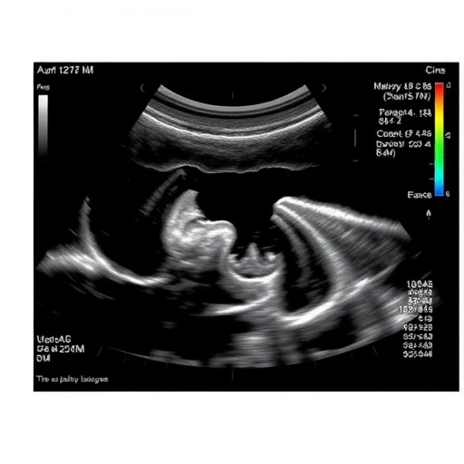
Transvaginal ultrasound can successfully rule out endometrial cancer in most Black women, even when uterine fibroids make the scan more difficult to complete, according to a new cohort study published in JAMA Network Open. The findings challenge concerns that lower ultrasound completion rates in Black women necessarily translate into a reduced ability to detect or exclude disease. Instead, the study suggests that the main obstacle is technical image quality, particularly when fibroids obscure the endometrium, while the test’s diagnostic performance remains strong when adequate images are obtained.
The study examined transvaginal sonography, an imaging technique in which a slender ultrasound probe is placed inside the vagina to produce high-resolution images of the uterus, cervix, ovaries, and endometrial lining. Unlike abdominal ultrasound, the transvaginal approach places the probe closer to the pelvic organs, allowing clinicians to evaluate the thickness and structure of the endometrium. This measurement is commonly used when assessing patients for possible endometrial cancer, particularly when abnormal uterine bleeding or other clinical concerns are present.
Researchers found that ultrasound examinations were completed less often among Black women than among women from other racial and ethnic groups. The difference was primarily associated with uterine fibroids, benign growths that develop from the muscular wall of the uterus. Fibroids are common and can distort the uterine cavity, change the position of the uterus, or create acoustic shadows that interfere with the ultrasound beam. When the endometrium cannot be visualized clearly from one edge to the other, the examination may be classified as incomplete, even if other pelvic structures are visible.
Despite this limitation, transvaginal sonography achieved a 76% completion rate among Black women in the cohort. More importantly, when the examination was technically adequate, the test retained an excellent ability to exclude endometrial cancer, whether or not fibroids were present. In clinical terms, this means that a sufficiently clear scan showing a reassuring endometrial appearance continued to provide meaningful evidence against cancer. Fibroids made imaging harder, but they did not appear to undermine the test’s underlying ability to identify women at low risk when the endometrium could be assessed.
The distinction between test completion and test accuracy is central to interpreting the findings. A study can fail to produce a usable image in some patients without becoming intrinsically less reliable in those who receive an interpretable examination. Completion rate measures whether the relevant anatomy can be visualized adequately; diagnostic performance measures how well the findings correspond with the presence or absence of disease. By separating these two outcomes, the researchers show that a lower completion rate should not automatically be interpreted as evidence that ultrasound is less effective for Black patients.
The results also illustrate how biological variation and imaging technology can interact with health disparities. Fibroids may affect women of all racial and ethnic backgrounds, but their prevalence, number, size, and clinical impact can vary across populations. A racial difference in completion rates therefore does not establish that race itself changes the performance of ultrasound. Instead, it may reflect differences in the frequency or characteristics of conditions that affect visualization, as well as differences in access to specialized imaging, follow-up procedures, and experienced sonographers.
For clinicians, the findings support a more nuanced approach when an ultrasound examination is incomplete. An obscured endometrium should not be treated as equivalent to a normal result, because the inability to see the lining clearly limits the examination’s ability to assess disease. Depending on the patient’s symptoms, risk factors, and clinical history, physicians may consider additional evaluation, such as repeat imaging, saline infusion sonography, hysteroscopy, or endometrial tissue sampling. The appropriate next step remains a clinical decision rather than a conclusion that can be made from race or fibroid status alone.
The study is particularly relevant because endometrial cancer is often diagnosed after symptoms have appeared, and delays in evaluating abnormal bleeding can have serious consequences. A test that can reliably identify patients at low risk may help reduce unnecessary invasive procedures, but only when its technical limitations are recognized. The findings suggest that improving image acquisition and developing clearer pathways for incomplete examinations could be more productive than abandoning transvaginal ultrasound for groups with higher rates of fibroids.
The researchers’ conclusion is therefore both reassuring and cautionary: transvaginal sonography remains a useful tool for excluding endometrial cancer in Black women, including those with fibroids, but clinicians must distinguish a confidently visualized endometrium from an examination in which the lining is hidden. Future research may help determine whether advances in ultrasound equipment, standardized scanning protocols, artificial intelligence-assisted image interpretation, or more consistent follow-up strategies can raise completion rates and further narrow disparities in gynecologic cancer evaluation.
Web References: https://doi.org/10.1001/jamanetworkopen.2026.27283
Cite Scienmag News
Nathaniel Bowman. (August 4, 2026). Study finds racial differences in ultrasound evaluation of suspected endometrial cancer. Scienmag. https://scienmag.com/study-finds-racial-differences-in-ultrasound-evaluation-of-suspected-endometrial-cancer/
Nathaniel Bowman. "Study finds racial differences in ultrasound evaluation of suspected endometrial cancer." Scienmag, 4 August 2026, https://scienmag.com/study-finds-racial-differences-in-ultrasound-evaluation-of-suspected-endometrial-cancer/. Accessed 5 September 2026.
Nathaniel Bowman. "Study finds racial differences in ultrasound evaluation of suspected endometrial cancer." Scienmag. August 4, 2026. https://scienmag.com/study-finds-racial-differences-in-ultrasound-evaluation-of-suspected-endometrial-cancer/


