


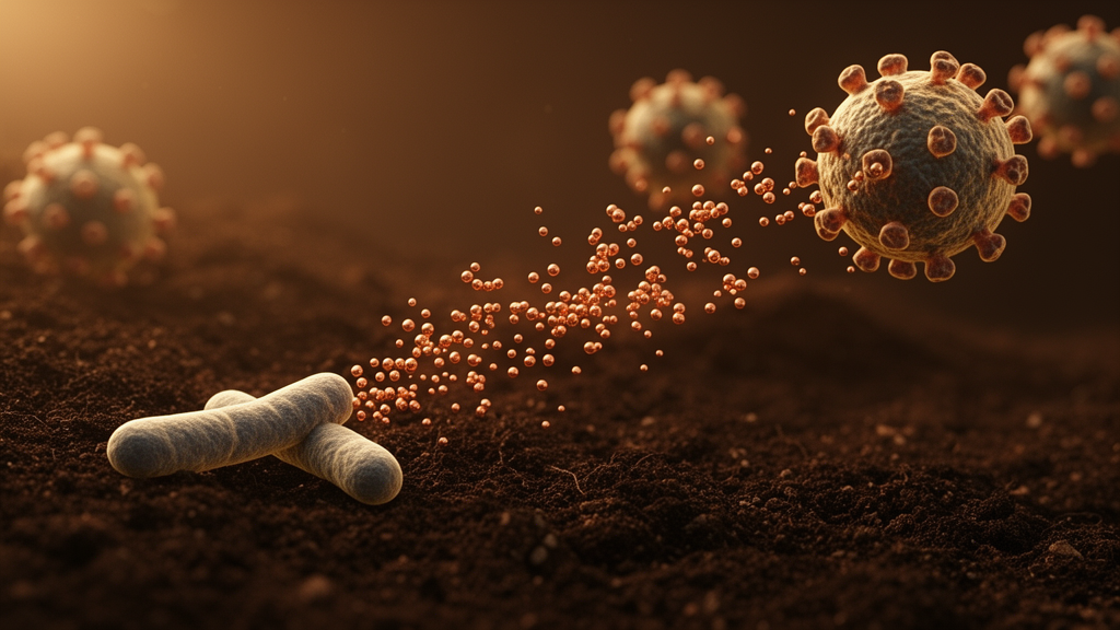
Researchers in Morocco and France have found a way to turn an ordinary soil bacterium into a nanoscale pharmacy, coaxing it to produce copper oxide nanoparticles that kill some of the most feared drug-resistant pathogens on the planet. The new study, published in Frontiers in Pharmacology, describes a green synthesis route in which the actinobacterium Streptomyces thinghirensis (DSM 41919T) acts as both reducing and stabilizing agent, yielding uniformly tiny spherical particles with potent antibacterial and antibiofilm activity against vancomycin-resistant Enterococcus faecium, methicillin-resistant Staphylococcus aureus and ceftazidime-resistant Pseudomonas aeruginosa. The work arrives at a moment when the World Health Organization has placed all three of these pathogens on its updated Bacterial Priority Pathogens List as high-priority threats, underscoring how urgently new weapons are needed.
The logic of the approach rests on a simple contrast. Conventional antibiotic pipelines are slow, expensive and increasingly outpaced by bacterial evolution, while chemical methods for manufacturing nanoparticles rely on toxic solvents and costly instrumentation. Biological synthesis, by contrast, is inexpensive, clean and operates at ambient conditions. The idea dates back to 1980, when Beveridge and Murray first showed that Bacillus subtilis could build nanosized gold. Since then, bacteria, plants, yeasts, fungi and actinomycetes have all been recruited as living nanofactories. Streptomyces species, famous for producing roughly half of all clinically used antibiotics, have a particular advantage: their rich secondary metabolism provides a natural toolkit of biomolecules that can reduce metal ions and cap the resulting particles, preventing them from clumping together.
In the new work, the team collected the cell-free supernatant of S. thinghirensis and mixed it in equal volume with a freshly prepared solution of copper sulfate pentahydrate at ten millimolar concentration. The mixture was incubated at 30 degrees Celsius in darkness for up to 120 hours. As the reaction proceeded, the solution changed color from light blue to greenish brown, the first visible hint that copper ions were being transformed into copper oxide nanoparticles. Ultraviolet-visible spectroscopy tracked the reaction kinetically: absorption in the 210 to 220 nanometer region rose steadily over the incubation period, reflecting progressive complexation of the bacterial secondary metabolites during the capping process, while the characteristic broad absorption shoulder of monoclinic copper oxide between 250 and 300 nanometers emerged clearly after 24 hours and peaked at 120 hours, which the researchers selected as the optimum reaction time.
A battery of analytical techniques then confirmed the identity, purity and dimensions of the product. X-ray diffraction revealed peaks at 38.04, 44.16, 62.05, 76.80 and 81.14 degrees, matching the (111), (200), (-202), (022) and (222) planes of monoclinic CuO and agreeing closely with the standard reference data, with no trace of metallic copper or cuprous oxide impurity phases. Fourier transform infrared spectroscopy identified the chemical fingerprints of the capping layer: strong bands near 490 and 563 per centimeter corresponding to copper-oxygen vibrations, alongside signals from amines, peptide carbonyls and hydroxyl groups that demonstrate how the bacterial metabolites wrap around the growing crystals. Energy-dispersive X-ray analysis showed the surface dominated by copper at roughly 48 percent and oxygen at about 28 percent, with smaller contributions of phosphorus, carbon, calcium and potassium inherited from the actinobacterial metabolites.
Transmission electron microscopy provided the most striking image of the material: well-dispersed, quasi-spherical particles averaging about 9.8 nanometers in diameter, with sizes ranging from 6 to 16 nanometers. That is considerably smaller than many copper oxide particles previously made with plant extracts, such as the 40 to 80 nanometer particles produced from papaya leaf extracts or the roughly 15 nanometer particles from Calotropis procera latex. Dynamic light scattering, which measures the hydrodynamic diameter of particles in solution rather than the dry inorganic core, gave a larger average of 54.2 nanometers with a bimodal distribution, a discrepancy the authors attribute to the organic capping ligands and hydration shell that DLS counts but TEM does not. A zeta potential of minus 13.5 millivolts indicated moderate colloidal stability, and selected-area electron diffraction confirmed crystallinity with lattice spacings matching the diffraction data.
With the material thoroughly characterized, the researchers turned to the microbes. They tested the particles, dubbed ST@CuONPs, against six strains: vancomycin-sensitive E. faecalis ATCC 29212, vancomycin-resistant E. faecium ATCC 700221 carrying the vanA gene, methicillin-susceptible S. aureus ATCC 29213, methicillin-resistant S. aureus NCTC 12493, ceftazidime-sensitive P. aeruginosa ATCC 27853 and ceftazidime-resistant P. aeruginosa ATCC BAA-2108. In disk diffusion assays, the lowest nanoparticle dose produced no inhibition, but activity rose steeply and dose-dependently at higher loads. At 13.5 micrograms per disk, the particles generated inhibition zones of 29 millimeters against E. faecalis, 30 millimeters against susceptible S. aureus and 23 millimeters against the resistant S. aureus strain, all statistically superior to the standard antibiotic disks used as comparators.
The most dramatic results came from the pathogens that no longer respond to conventional therapy. The vancomycin-resistant E. faecium strain, which showed a zero-millimeter zone against the reference antibiotic, was highly susceptible to the nanoparticles, yielding a 29.17-millimeter inhibition zone. The ceftazidime-resistant Pseudomonas strain also proved susceptible, with a 10-millimeter zone, although the resistant Gram-negative pathogen was clearly harder to kill than its Gram-positive counterparts. Broth microdilution confirmed this pattern quantitatively: minimum inhibitory concentrations were 12.5 micrograms per milliliter for the sensitive E. faecalis and S. aureus strains, 25 micrograms per milliliter for the resistant E. faecium and S. aureus, and 100 micrograms per milliliter for both Pseudomonas strains. The Gram-negative envelope, with its outer lipid-rich membrane, appears to blunt the copper attack, whereas copper’s affinity for proteins makes the thick peptidoglycan walls of Gram-positive bacteria, roughly 30 nanometers thick compared with 3 to 4 nanometers in Gram-negative species, especially vulnerable.
Perhaps the most clinically significant finding concerns biofilms, the slimy, surface-attached bacterial communities that shield pathogens from both antibiotics and immune cells and serve as hotspots for the exchange of resistance genes. Using crystal violet staining to quantify preformed biofilms, the team showed that the nanoparticles dismantled these structures in a concentration-dependent manner. Against the vancomycin-resistant E. faecium strain, biofilm mass fell by 64 percent at a quarter of the MIC, by 83 percent at half the MIC and by 89 percent at the full MIC. The susceptible E. faecalis strain was even more responsive, retaining only 8 percent of its biofilm at the full MIC, a 92 percent eradication. Even the recalcitrant Pseudomonas biofilms were significantly reduced, by 68 and 58 percent at full MIC for the sensitive and resistant strains respectively. The authors propose that the bactericidal mechanism operates on several fronts at once: generation of reactive oxygen species, disruption of bacterial cell membranes, DNA damage and inhibition of protein synthesis, a multipronged assault that is difficult for bacteria to counter with single-gene resistance mechanisms.
The study is not without caveats. The authors acknowledge that they lacked a concurrent, internal cell-free supernatant control within the same experimental runs, although their earlier work established only a modest baseline effect for the supernatant alone, 12 to 14 millimeter zones, far short of the much larger zones observed here with the nanoparticles. They also frame the antibacterial mechanism as a hypothetical framework drawn from prior literature rather than a fully resolved pathway, and the moderate zeta potential suggests the colloidal stability could be improved. Still, the eco-friendly synthesis, which avoids toxic reagents entirely and uses a cheap, abundant metal instead of gold, platinum or silver, makes the route economically attractive, particularly for developing countries where antimicrobial resistance exacts its heaviest toll.
For now, the particles remain a laboratory-scale proof of concept rather than a bedside therapy. Toxicity testing, pharmacokinetics, formulation and large-scale manufacturing all lie ahead. But the demonstration that a single soil-dwelling Streptomyces strain can fabricate near-ten-nanometer copper oxide particles capable of shrinking vancomycin-resistant biofilms by nearly 90 percent offers a tantalizing glimpse of where the fight against superbugs may go next: not toward yet another tweak on an old antibiotic molecule, but toward materials engineered, or rather, grown, by biology itself.
Subject of Research: Green synthesis of copper oxide nanoparticles by Streptomyces thinghirensis and their activity against multidrug-resistant bacteria
Article Title: Green synthesis of copper oxide nanoparticles ST@CuONPs by Streptomyces thinghirensis: current treatment path against multidrug-resistant bacteria
Article References: Green synthesis of copper oxide nanoparticles ST@CuONPs by Streptomyces thinghirensis: current treatment path against multidrug-resistant bacteria. (n.d.). https://doi.org/10.3389/fphar.2026.1904965
Image Credits: AI Generated
DOI: 10.3389/fphar.2026.1904965
Keywords: copper oxide nanoparticles, green synthesis, Streptomyces thinghirensis, antimicrobial resistance, multidrug-resistant bacteria, biofilms, nanotechnology, vancomycin-resistant Enterococcus faecium, MRSA, Pseudomonas aeruginosa, actinomycetes, antibiofilm activity
Cite Scienmag News
Ophelia Keating. (October 15, 2026). Soil Bacteria Grow Tiny Copper Particles That Attack Drug-Resistant Superbugs. Scienmag. https://scienmag.com/soil-bacteria-grow-tiny-copper-particles-that-attack-drug-resistant-superbugs/
Ophelia Keating. "Soil Bacteria Grow Tiny Copper Particles That Attack Drug-Resistant Superbugs." Scienmag, 15 October 2026, https://scienmag.com/soil-bacteria-grow-tiny-copper-particles-that-attack-drug-resistant-superbugs/. Accessed 15 October 2026.
Ophelia Keating. "Soil Bacteria Grow Tiny Copper Particles That Attack Drug-Resistant Superbugs." Scienmag. October 15, 2026. https://scienmag.com/soil-bacteria-grow-tiny-copper-particles-that-attack-drug-resistant-superbugs/


