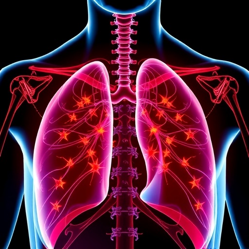
In a groundbreaking study poised to reshape clinical management strategies, researchers have unveiled compelling evidence linking skeletal muscle loss with overall survival outcomes in patients battling limited-stage small-cell lung cancer (LD-SCLC) undergoing concurrent chemoradiotherapy (CCRT). This investigation, recently published in the prestigious journal BMC Cancer, highlights the critical prognostic significance of muscle degradation measured during treatment, propelling a deeper understanding of cachexia’s role in oncologic therapies.
Skeletal muscle mass, a biological determinant long appreciated for its influence on general health and physical resilience, emerges as a vital biomarker in this context. Utilizing advanced body composition analysis at precise vertebral landmarks, the study meticulously quantified changes in the skeletal muscle index (SMI)—particularly at the third lumbar vertebral level (L3) and fourth thoracic vertebral level (T4)—throughout the rigorous course of CCRT. This methodological precision enabled the delineation of nuanced muscular shifts that correlate robustly with survival probabilities.
The research cohort comprised 55 patients diagnosed with LD-SCLC, a highly aggressive form of lung cancer typified by rapid proliferation and early metastatic potential. These patients underwent comprehensive chemoradiotherapy, a formidable therapeutic regimen combining systemic chemotherapy with localized radiation aimed at maximizing tumor control. Despite the aggressive treatment intent, the investigation revealed that nearly half of the participants displayed low muscle mass at baseline according to culturally and population-specific Korean criteria, underscoring preexisting vulnerabilities that may predispose patients to adverse outcomes.
Intriguingly, longitudinal assessment of body composition exposed a significant median decline of approximately 5.8 cm²/m² in L3 SMI post-treatment, a magnitude that was statistically significant (p < 0.001). Contrastingly, changes in T4 SMI, body mass index (BMI), and overall body weight did not achieve statistical significance, suggesting that lumbar skeletal muscle may serve as a more sensitive and prognostically relevant indicator than more conventional metrics during oncologic treatment.
Investigators pursued rigorous multivariate analyses to decipher the independent predictive power of muscle loss relative to established clinical factors. The Eastern Cooperative Oncology Group (ECOG) performance status—a validated measure of patient functional capacity—emerged as a dominant prognostic marker, with patients exhibiting ECOG status 2 facing markedly increased mortality risk. Critically, when adjusting for ECOG performance, the decrement in L3 SMI maintained its statistical significance, revealing a more than twofold elevation in mortality hazard (hazard ratio 2.172, p = 0.027). This interplay suggests that skeletal muscle wasting may potentiate the detrimental effect of diminished functional status.
The pathophysiological ramifications of these findings are profound. Skeletal muscle loss, frequently manifesting as cancer cachexia, contributes to systemic inflammation, metabolic derangements, and compromised immune competence. These alterations not only diminish tolerance to chemotherapy and radiotherapy but may exacerbate tumor progression through complex biochemical and molecular pathways. Hence, the muscle index changes observed are not merely numeric data points but reflect broader biological deterioration impacting patient survival trajectories.
Prior research offers limited consensus regarding the prognostic potency of tissue depletion at differing vertebral levels. The preferential sensitivity of L3 SMI identified in this study reinforces the utility of lumbar region imaging biomarkers in oncology. This is likely attributable to the consistent representation of muscle groups at L3, which correspond closely with whole-body musculature and metabolic health, in contrast to thoracic measurements that may be confounded by respiratory musculature and anatomical variability.
Moreover, the study’s retrospective design provides a pragmatic evaluation of real-world clinical scenarios but invites future prospective investigations. Interventional trials exploring nutritional supplementation, resistance exercise programs, and pharmacologic agents targeting cachexia biology could elucidate strategies to attenuate muscle loss during CCRT. The ultimate goal being enhanced survival through preservation or restoration of skeletal muscle integrity.
The implications extend beyond direct patient care to inform imaging protocols and multidisciplinary treatment planning. Regular, standardized assessment of skeletal muscle mass via computed tomography or magnetic resonance imaging at baseline and during treatment could become an integral component of oncologic monitoring. Early identification of patients exhibiting rapid muscle decline may enable tailored supportive interventions, optimizing drug dosing and mitigating treatment-related toxicity.
Understanding the intersection between muscle biology and oncologic therapeutics further prompts exploration into molecular mechanisms underpinning this association. Inflammatory cytokines such as TNF-alpha and IL-6, ubiquitin-proteasome pathways, and myostatin signaling represent potential molecular targets implicated in muscle catabolism amidst cancer treatment. Integrating biomarker research with clinical data promises to unravel therapeutic vulnerabilities and inspire novel adjunct treatments.
The study also highlights the importance of performance status evaluation in prognostication, reinforcing jejune but crucial clinical assessments. Stratification by ECOG performance enables holistic appraisal encompassing physical function, symptom burden, and treatment tolerance, which, when combined with quantitative muscle measurements, offers a nuanced approach to patient risk assessment.
This research underscores the necessity for interdisciplinary collaboration, converging oncology, radiology, nutrition, physical therapy, and molecular biology expertise to enhance patient outcomes. The dynamic monitoring of skeletal muscle during intensive treatment regimens epitomizes precision medicine principles, tailoring supportive care based on individualized risk profiles.
Ultimately, the evidence presented advocates for a paradigm shift in managing LD-SCLC, where skeletal muscle conservation is recognized not only as a quality-of-life objective but as a determinant of survival. The integration of muscle mass assessment into clinical guidelines holds promise to refine prognostic accuracy, personalize therapeutic interventions, and improve life expectancy among patients confronting this formidable malignancy.
As lung cancer remains a leading cause of cancer-related mortality globally, innovations such as these represent pivotal advances. Harnessing the predictive capability of skeletal muscle status may redefine treatment algorithms, ushering in an era where multimodal care encounters biologically informed metrics to optimize efficacy and patient resilience.
In conclusion, skeletal muscle loss emerges as a critical biomarker in small-cell lung cancer patients receiving chemoradiotherapy, intricately linked with overall survival outcomes. This research accentuates the need for vigilant assessment and strategic intervention addressing muscle depletion, heralding significant implications for clinical practice, patient management, and future research directions.
Subject of Research: Skeletal muscle loss and its prognostic significance in limited-stage small-cell lung cancer patients undergoing concurrent chemoradiotherapy.
Article Title: Skeletal muscle loss and associated clinical outcomes in patients with small-cell lung cancer receiving concurrent chemoradiotherapy.
Article References:
Park, S.E., Hwang, I.G. & Choi, J.H. Skeletal muscle loss and associated clinical outcomes in patients with small-cell lung cancer receiving concurrent chemoradiotherapy. BMC Cancer 25, 1772 (2025). https://doi.org/10.1186/s12885-025-15141-5
Image Credits: Scienmag.com
DOI: 10.1186/s12885-025-15141-5 (Published 17 November 2025)


