


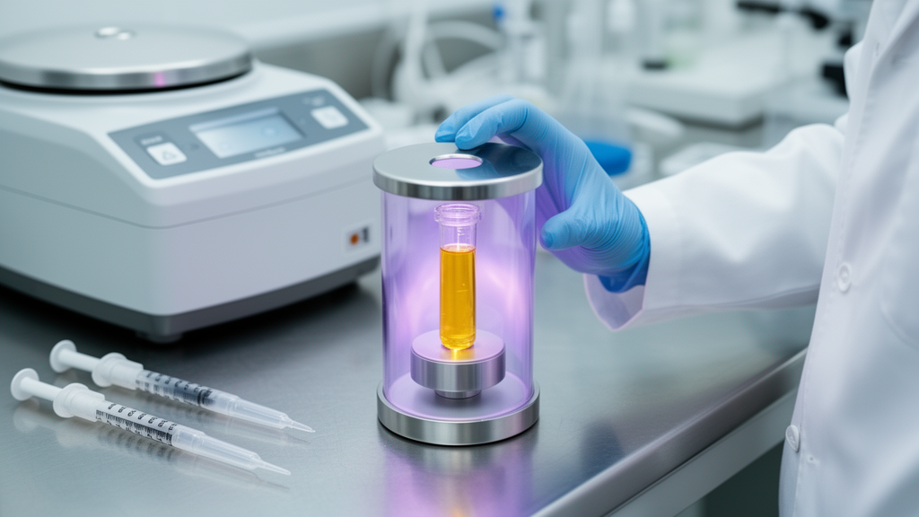
Researchers have built a device that turns an ordinary clinical blood collection tube into its own plasma reactor, activating platelet-rich plasma without ever opening the vial. The system, described as a vial wall-based dielectric barrier discharge (DBD) plasma activation device, uses the tube's own wall as the dielectric barrier that cold atmospheric plasma requires. In a proof-of-concept translational study, plasma-activated platelet-rich plasma (PA-PRP) prepared this way showed stronger platelet activation and higher growth factor concentrations than conventional PRP, reduced cartilage damage in an osteoarthritic mouse model, and was delivered to three patients with knee osteoarthritis in a small split-body feasibility study with no recorded adverse events.
The engineering insight at the heart of the device is deceptively simple. Standard blood collection tubes already possess high dielectric strength, cylindrical symmetry, hermetic sealing, and pre-sterilization, which map directly onto the requirements of dielectric barrier discharge generation. Rather than exposing liquid to an open plasma jet, the team placed high-voltage and grounded electrodes on the outer wall of a sealed tube.
When the pulsed power supply is switched on, a blue filamentary corona discharge ignites in the headspace above the liquid inside the tube, and the plasma field activates the sample through the wall. The container is never breached, so the original sterility of the sample is preserved throughout the process. Electrical characterization showed the prototype operating at a minimum sustaining input power of 3 watts, with peak output voltage and current of approximately 6.08 kilovolts and 7.45 milliamperes at a frequency of 10 kilohertz.
Lissajous figure analysis with a 0.47-microfarad sampling capacitor yielded an output power of 1.58 plus or minus 0.18 watts, corresponding to a discharge efficiency of about 52.57 percent. Notably, this efficiency remained nearly identical under the optimized activation conditions, suggesting that the discharge behavior is governed primarily by the fixed geometry and material properties of the tube wall rather than by operator technique. The device also produced stable discharge in blood collection tubes from five different commercial brands, despite variation in wall material, thickness, and dielectric constant.
Finding a safe operating window was a central task. The researchers tested combinations of input power, duty cycle, and activation duration, tracking product temperature, pH, and reactive oxygen species concentration. Longer or more intense activation raised the temperature and increased ROS levels, while extending activation beyond 30 seconds dropped the pH sharply beyond the buffering capacity of the plasma.
They settled on 6 watts of input power, a 1 percent duty cycle, and 15 seconds of activation, which kept the final product at 36.7 plus or minus 1.5 degrees Celsius and a pH of 7.307 plus or minus 0.031, both within physiological limits. Across 50 consecutive activations, the coefficient of variation in ROS concentration was only 4.53 percent, an initial estimate of reproducibility during continuous operation. The contamination advantage of the sealed design was among the most striking results.
PA-PRP activated in the sealed tube showed no bacterial colonies after 24-hour incubation on LB agar, and no colonies of eukaryotic microorganisms or anaerobic bacteria on appropriate culture media. Endotoxin in all tube-sealed samples was below the detection limit of a chromogenic limulus amebocyte lysate assay. In contrast, PA-PRP activated by a conventional open plasma jet, which required breaking the tube seal, produced obvious eukaryotic microbial colonies on Sabouraud dextrose agar and contained detectable trace endotoxin ranging from 200 to 1500 picograms per milliliter across replicates.
For intra-articular injection, where iatrogenic infection can cause septic arthritis and irreversible joint damage, this reduced contamination risk is a meaningful safety gain, although the authors caution that pharmaceutical-grade sterility validation under good manufacturing practice standards remains to be performed. Biologically, plasma activation appeared to wake the platelets up. Immunofluorescence staining showed that 43.04 plus or minus 9.67 percent of CD41-positive platelets in PA-PRP expressed the activation marker CD63, significantly more than in untreated PRP.
Enzyme-linked immunosorbent assays on paired aliquots found higher concentrations of TGF-beta, total PDGF, and IGF-1 after activation, and a cytokine microarray revealed elevated levels of additional factors including PDGF-BB, VEGF, IL-10, IL-4, IL-1 receptor antagonist, and insulin-like growth factor binding proteins. In cell culture, medium supplemented with PA-PRP shifted macrophage marker expression away from the inflammatory M1 marker iNOS and toward the M2 markers CD206 and arginase 1, and adding 10 millimolar of the ROS scavenger N-acetylcysteine attenuated these changes, supporting a role for plasma-derived reactive species. Qualitative electron paramagnetic resonance spin trapping suggested singlet oxygen rather than hydroxyl radical signals, though the specific responsible species and their persistence were not identified.
In a mouse model of osteoarthritis induced by destabilization of the medial meniscus, weekly intra-articular injections of PA-PRP were associated with better gait outcomes than conventional PRP. The composite gait recovery index reached 97.69 plus or minus 1.36 percent in the PA-PRP group versus 86.35 plus or minus 4.35 percent with PRP, with differences in stride length, stride width, and paw rotation angle surviving statistical correction. Histological OARSI scoring showed lower cartilage degeneration scores with PA-PRP, along with reduced levels of the matrix-degrading enzymes MMP9, MMP3, and MMP13.
The authors are careful to note that gait measures are not direct measures of pain, that the study used only five mice per group without a prospective power calculation, and that these findings are hypothesis-generating rather than definitive efficacy proof. Human osteoarthritic cartilage explants, harvested from discarded knee tissue of five patients undergoing joint replacement, provided convergent evidence. After 14 days in culture, explants treated with PA-PRP showed significantly higher normalized Safranin O staining intensity, a marker of proteoglycan content, and higher Alcian blue staining, which labels acidic glycosaminoglycans, than explants treated with PRP or control medium.
This suggested that the ex vivo model preserved the pattern seen in mice and supported further investigation of possible chondroprotective activity, while remaining a laboratory observation under defined culture conditions rather than a clinical result. The clinical component was deliberately modest: a randomized, single-blind, split-body study in three patients with bilateral mild-to-moderate knee osteoarthritis, each receiving PA-PRP in one knee and conventional PRP in the other, three weekly injections per knee. At week 15, both knees had improved, with descriptive mean VAS pain reductions of 72.20 plus or minus 25.47 percent for PA-PRP versus 44.40 plus or minus 19.23 percent for PRP, WOMAC improvements of 49.00 versus 27.67 percent, and larger effusion volume reductions in the PA-PRP knees.
No adverse events were recorded at any scheduled visit. The authors explicitly present these numbers without inferential testing, as feasibility observations to guide sample size planning and endpoint selection for an adequately powered randomized controlled trial, which they report is now under preparation. Limitations frame the promise.
The final PRP platelet recovery, leukocyte composition, and residual erythrocyte content were not systematically quantified, so activation effects could not be fully separated from variability in starting material. No head-to-head comparison was made with chemically activated PRP using thrombin or calcium chloride, and the prototype, at roughly US$4000 with separate components, awaits integration into a compact point-of-care format projected to cost under US$1000. Still, the core achievement stands: a fully enclosed, standardized, operator-independent method of plasma activation that keeps the therapeutic sealed from vein to joint, addressing the sterility, reproducibility, and quality control barriers that have kept plasma medicine largely confined to the laboratory.
Subject of Research: A sealed-tube dielectric barrier discharge plasma activation device for preparing plasma-activated platelet-rich plasma to treat knee osteoarthritis
Article Title: Clinical validation of a vial wall-based dielectric barrier discharge plasma activation device for plasma-activated platelet-rich plasma preparation in osteoarthritis pain management
Article References: Wang, S., Li, Z., Gong, Z., Deng, Z., Jin, Z., Pang, B., Yao, J., Zhang, S., Zhu, W., Han, J., Dai, Y., Wan, L., Wan, J., Liu, M., Tang, S., Jiang, L., & Wang, X. (2026). Clinical validation of a vial wall-based dielectric barrier discharge plasma activation device for plasma-activated platelet-rich plasma preparation in osteoarthritis pain management. Materials Today Bio, 41, Article 103766. https://doi.org/10.1016/j.mtbio.2026.103766
Image Credits: AI Generated
DOI: 10.1016/j.mtbio.2026.103766
Keywords: cold atmospheric plasma, dielectric barrier discharge, platelet-rich plasma, osteoarthritis, growth factors, contamination risk, macrophage polarization, cartilage explants, mouse model, clinical feasibility study, reactive oxygen species, biomedical device
Cite Scienmag News
Denise Maddox. (October 26, 2026). Research report: Clinical validation of a vial wall-based dielectric barrier discharge plasma activation device for plasma-activated platelet-rich plasma preparation in osteoarthritis pain management. Scienmag. https://scienmag.com/research-report-clinical-validation-of-a-vial-wall-based-dielectric-barrier-discharge-plasma-activation-device-for-plasma-activated-platelet-rich-plasma-preparation-in-osteoarthritis-pain-management/
Denise Maddox. "Research report: Clinical validation of a vial wall-based dielectric barrier discharge plasma activation device for plasma-activated platelet-rich plasma preparation in osteoarthritis pain management." Scienmag, 26 October 2026, https://scienmag.com/research-report-clinical-validation-of-a-vial-wall-based-dielectric-barrier-discharge-plasma-activation-device-for-plasma-activated-platelet-rich-plasma-preparation-in-osteoarthritis-pain-management/. Accessed 26 October 2026.
Denise Maddox. "Research report: Clinical validation of a vial wall-based dielectric barrier discharge plasma activation device for plasma-activated platelet-rich plasma preparation in osteoarthritis pain management." Scienmag. October 26, 2026. https://scienmag.com/research-report-clinical-validation-of-a-vial-wall-based-dielectric-barrier-discharge-plasma-activation-device-for-plasma-activated-platelet-rich-plasma-preparation-in-osteoarthritis-pain-management/


