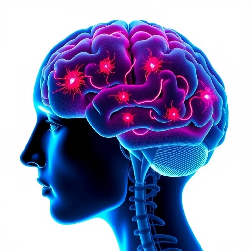
In a groundbreaking study poised to reshape our understanding of sports-related brain injuries, researchers have unveiled compelling evidence that repeated head trauma alone can induce significant neuronal loss and inflammation in young athletes. This research not only sheds light on the biological consequences of repeated head impacts but also challenges traditional assumptions about the timeline and mechanisms underlying neurodegenerative diseases commonly linked with such injuries.
The central focus of the investigation was on excitatory neurons within the cerebral cortex, particularly those residing in layers 2 and 3, which play essential roles in cognitive functions and neural circuit integration. Using advanced single-nucleus RNA sequencing (snRNA-seq) techniques, the team meticulously scrutinized the transcriptomic landscape of neurons from individuals exposed to repetitive head impacts, including those with chronic traumatic encephalopathy (CTE) and those with repetitive head injury (RHI) but no formal CTE diagnosis. Their findings revealed pronounced disruptions in gene expression profiles linked to synaptic function and cellular adhesion pathways.
Interestingly, while the alterations in excitatory neurons were substantial, inhibitory neurons demonstrated comparatively fewer transcriptional changes. Approximately 47% of excitatory neuron differentially expressed genes (DEGs) were common between RHI and CTE cases when compared to control samples, suggesting that the initial exposure to repetitive head trauma drives the majority of transcriptomic disturbances. These changes encompass critical genes involved in synaptic transmission, including SYN3, SNAP91, NRG1, and HSP12A1—the latter belonging to the heat shock protein family known for its neuroprotective roles.
Complementing these molecular insights, the researchers delved into the cell-specific effects of trauma exposure. A striking observation was the selective vulnerability and subsequent loss of a particular subtype of excitatory neurons characterized by the co-expression of the markers CUX2 and LAMP5. These neurons, localized primarily at the depth of cortical sulci, exhibited a marked reduction in individuals with a history of repetitive head injuries—irrespective of CTE diagnosis. Quantitative analysis unveiled a reduction of approximately 56% fewer CUX2+LAMP5+ neurons in those exposed to RHI compared with age-matched controls.
This neuronal depletion showed a clear correlation with years of football play or other contact sports participation, underscoring the cumulative detrimental effect of chronic repetitive impacts. Spatial mapping of these neurons through in situ hybridization confirmed their diminished density specifically at sulcal depths rather than at gyral crests, suggesting localized susceptibility likely attributable to biomechanical forces concentrated in these cortical regions. Furthermore, this vulnerability appears exclusive to the CUX2+LAMP5+ subset; other neighboring excitatory neurons expressing CUX2 alone remained unaffected, highlighting a unique susceptibility profile.
Expanding upon histological validation, the study employed Nissl staining across a broader cohort of young athletes ranging from zero to 28 years of football exposure. Neuronal density in layers 2/3 of the sulcus declined significantly with increased years of playing contact sports, independent of the individual’s age at death. This layer- and region-specific neuronal loss corroborates the transcriptomic data and points toward early, subclinical brain changes precipitated by repetitive head impacts.
Crucially, the research found no association between neuronal loss and phosphorylated tau (p-tau) pathology, a hallmark protein aggregation commonly implicated in neurodegenerative disorders such as CTE and Alzheimer’s disease. This decoupling of neuronal depletion from p-tau deposition suggests that neurodegeneration may be initiated independently of—or even precede—classical pathological protein accumulation in the early stages following head trauma.
Beyond neurons, the study illuminated potential immunological consequences linked to repetitive head impacts. Analysis of microglial populations, the brain’s resident immune cells, revealed that homeostatic microglia expressing high levels of P2RY12 and IBA1 co-localize and correlate positively with neuronal densities in layers 2/3. This relationship points to a possible feedback mechanism where the loss of neurons may disturb microglial homeostasis or vice versa, potentially amplifying neuroinflammation and secondary injury cascades.
Taken together, these findings illustrate a mechanistic cascade where repeated head trauma initiates synaptic dysfunction and targeted neuronal loss within specific cortical layers, precipitating neuroinflammatory responses that may compound neural damage. Importantly, this process unfolds in the absence of overt tau pathology, reorienting the timeline of neurodegeneration in athletes exposed to head injuries and possibly explaining early cognitive and behavioral symptoms observed clinically.
The revelation that repetitive head trauma alone can drive significant neuronal loss challenges current diagnostic paradigms for sports-related brain injury syndromes. It underscores the urgent need for earlier detection methods and intervention strategies aimed at preserving neuronal populations before irreversible neurodegenerative changes ensue. Moreover, it highlights the critical importance of protective measures in contact sports, emphasizing cumulative exposure risks rather than solely the presence of overt clinical symptoms or neuropathological hallmarks.
Future research is warranted to unravel the exact molecular pathways mediating the selective susceptibility of excitatory CUX2+LAMP5+ neurons to mechanical insults and to clarify the role of microglial activity in either mitigating or exacerbating neuronal damage. Such insights could pave the way for therapeutic interventions targeting neuron-microglia interactions or synaptic stabilization to prevent or slow disease progression.
In addition, longitudinal studies tracking young athletes over time with integrated neuroimaging, biomarker profiling, and cognitive assessments would be instrumental in validating these findings and translating them into clinically actionable guidelines. Considering the profound public health implications of sports-related brain injuries, this research marks a pivotal step toward unraveling the complex interplay between mechanical trauma, neuronal integrity, and neuroinflammation.
As professional sports leagues, medical professionals, and policymakers grapple with the challenge of protecting athletes’ brain health, these data serve as a clarion call emphasizing that even in the absence of classic neurodegenerative pathology, the brain endures tangible, lasting harm from repetitive head trauma. Proactive measures, including revised concussion protocols, exposure limitations, and novel monitoring technologies, must be prioritized to safeguard athletes across all levels.
Ultimately, this study enriches our understanding of the biological sequelae following repeated head impact, providing a nuanced framework that recognizes early neuronal loss as a critical antecedent in the pathological continuum leading to dementia and other long-term neurological deficits. By highlighting the distinct molecular and cellular consequences of trauma before protein aggregation ensues, it lays the groundwork for redefining diagnostic criteria and therapeutic windows in sports-related neurodegenerative diseases.
Subject of Research: Neuronal and synaptic alterations caused by repeated head trauma in young athletes
Article Title: Repeated head trauma causes neuron loss and inflammation in young athletes
Article References:
Butler, M.L.M.D., Pervaiz, N., Breen, K. et al. Repeated head trauma causes neuron loss and inflammation in young athletes. Nature (2025). https://doi.org/10.1038/s41586-025-09534-6
Image Credits: AI Generated
