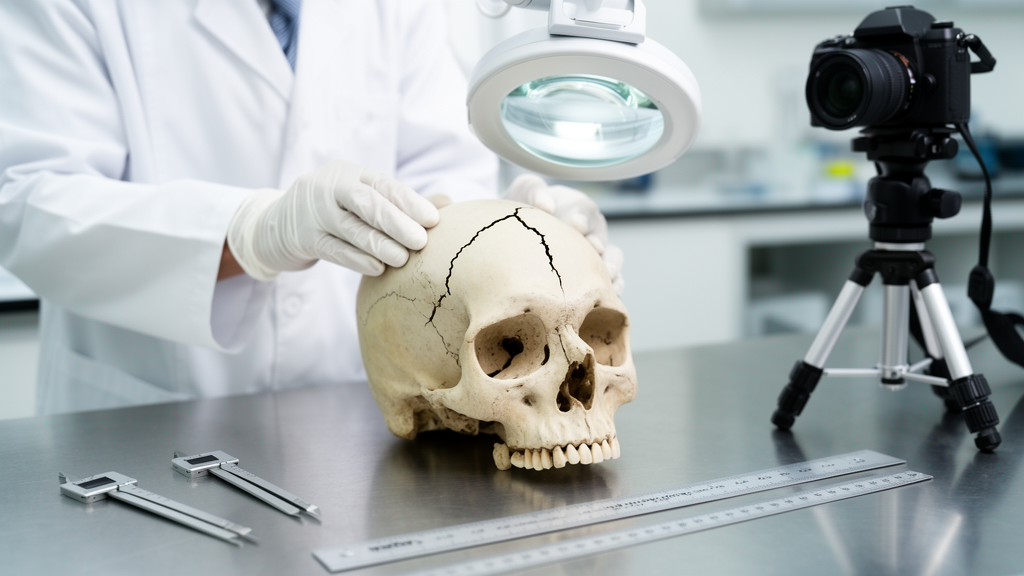
Skull fractures are among the most common findings in forensic autopsies, and interpreting them correctly can decide whether a death is understood as an accident, a suicide or a homicide. The authors stress that the assessment of skull fractures is a major issue in the daily work of forensic pathologists, precisely because bone damage from blunt force and ballistic trauma must be distinguished from lesions induced by heat or inflicted with sharp-edged weapons.
The review is deliberately practice-oriented. Rather than compiling epidemiological data or clinical treatment statistics, the authors focus on what a fracture actually looks like and what that appearance can tell an investigator. Most skull fractures arise from blunt traumatization in accidents, suicides or homicides, and the great majority result from direct impact with consecutive deformation of the neurocranium. Under specific conditions, however, the force may be applied far from the fracture site, which is one of the reasons many forensic pathologists feel uncertain when interpreting fatal craniocerebral trauma. The vast range of fracture types and their manifold appearances make this one of the trickiest areas of medico-legal practice.
The biomechanics behind the most frequent form, the linear fracture, follows an elegant geometric logic. The authors compare the human skull to a terrestrial globe covered by latitude and longitude lines. When a blunt impact strikes the dome-shaped skullcap, the bone is flattened and indented at the point of impact while bending outward in the periphery, where it is exposed to traction. This deformation lengthens the latitudinal lines, generating tensile stress that promotes one or more linear cracks. Notably, experimental work by Isa and colleagues, recorded on video, showed that the starting point of crack formation is typically peripheral to the site of force application, whereas Kroman and colleagues had previously argued that fractures radiate from the point of impact. Either way, one end of the fracture line is often in congruity with the impact site, which is usually marked by a corresponding scalp injury such as an abrasion, contusion or laceration.
For falls from a standing position, forensic tradition offers the hat brim line rule, introduced after Kratter’s 1919 description of a demarcation line for impact sites from falls to the ground. According to this rule, fall-related impact sites do not lie above the hat brim line when the fall occurred from standing to the ground, but the authors warn that it must not be applied to falls from a height, including stairs, or to traffic accidents. The statistics underscore how frequent such fractures are: in fatal falls from standing height, two thirds of victims sustain skull fractures, mostly single and linear, and a high proportion of the victims are under the influence of alcohol. Linear fractures can also be accompanied by epidural haematomas when an intersecting fracture lacerates branches of the middle meningeal artery, while the bleeding source of subdural haemorrhages typically lies away from any skull fracture.
Beyond the linear type, the review catalogues a gallery of special forms. Comminuted fractures, with more than two bone fragments, reflect a high-energy crushing impact, and the pieces can be displaced inward toward the brain as depressed fractures. Small-area impressions typically mirror the striking surface of the implement, such as a hammer head, a wrench or even a pistol grip, and in exceptional circumstances a depressed fracture may be caused by a kick to the head. At the base of the skull, hinge fractures run transversely from one side to the other across the floor of the middle cranial fossa, allowing the front and rear halves of the base to move independently after the skullcap is removed; they arise from one-sided lateral impacts or side-to-side compression and are frequently associated with blood aspiration because of their proximity to the sphenoid sinuses. Ring fractures encircling the foramen magnum, by contrast, result from indirect forces, either impaction of the upper cervical spine into the cranial cavity or traction at the craniocervical junction, as seen in high-speed collisions where the abruptly decelerated trunk leaves the head still moving.
Contrecoup fractures illustrate how force can injure bone far from where it was applied. Typically occurring after falls with impact to the occipital or temporal region, they appear as single or multiple fissures in the thin orbital roofs or the cribriform plate of the ethmoid, often with torn dura and, in short-term survivors, the formation of raccoon eyes. The review also covers elevated fractures, in which fragments are lifted outward on withdrawal of a penetrating blow; terraced fractures from angled blows producing concentric stepped depressions; and chop injuries from axes, swords, machetes and cleavers, characterized by linear notches, complete bone severance and edge chipping. Sequencing is addressed through Puppe’s rule, which holds that a second fracture ends at a pre-existing break, though the authors note possible exceptions, such as fractures passing through old craniotomies or recent burr holes.
Gunshot damage receives a detailed treatment grounded in wound ballistics. A distinction is drawn between direct interaction with the projectile and secondary fractures caused by the pressure rise from the temporary cavity forming within the confined space of the skull. High-speed video documentation of test shots to head models has shown that cracks radiating from the entrance hole appear only after the bullet has penetrated the bone, which then bulges outward and bursts; these radiating fractures can reach the opposite side of the skull before the bullet itself. In flat bone, a circular hole with internal bevelling usually indicates an entrance site, while exits show outward bevelled defects, although the authors caution that not all internally bevelled circular defects are gunshot entrances, citing a case produced by a thrust of an umbrella. Basal fractures away from the impact site are frequent in fatal handgun head shots, reported in the anterior cranial fossa in 82 percent of surveyed cases.
The review also addresses pitfalls and modern adjuncts. Subject-specific finite element head models have been developed to predict skull fractures in real accidents, with simulated fracture lines compared against post-mortem CT scans and autopsies, and CT-derived skull models can give courts a tangible three-dimensional impression of the injury pattern. Heat-induced fractures in burned bodies, post-mortem damage by animals, and the dating of fractures by healing stage, with all its uncertainties, round out the diagnostic landscape. In suspected child abuse, the authors emphasize that no fracture on its own can prove abusive causation, since even a fall from table height may cause a skull fracture.
The unifying message is a holistic one. Fracture assessment, following Berryman and colleagues, should rest on the triad of fracture characteristics, intrinsic factors and extrinsic factors, with reconstruction attempts incorporating scene findings, scalp injuries, entrance and exit wounds, post-mortem imaging and changes to the brain and meninges. Cranial fractures as such rarely threaten the victim’s life, even though they indicate substantial force on the bony braincase, but read correctly they can illuminate the course of events and the manner of death. For a field in which many practitioners admit uncertainty, this synthesis of morphology, biomechanics and reconstructive reasoning offers a practical compass for one of forensic medicine’s oldest and most consequential questions.
Subject of Research: A forensic review of skull fracture morphology and biomechanical trauma reconstruction
Article Title: Skull fractures in forensic autopsies: phenomenology and possibilities of biomechanical reconstruction*
Article References: Pollak, S., Große Perdekamp, M., & Thierauf-Emberger, A. (2026). Skull fractures in forensic autopsies: phenomenology and possibilities of biomechanical reconstruction*. International Journal of Legal Medicine. https://doi.org/10.1007/s00414-026-04044-0
Image Credits: AI Generated
DOI: 10.1007/s00414-026-04044-0
Keywords: forensic pathology, skull fractures, biomechanics, autopsy, gunshot wounds, blunt trauma, ring fracture, hinge fracture, Puppe's rule, post-mortem CT, trauma reconstruction, legal medicine
Cite Scienmag News
Ophelia Keating. (October 29, 2026). Reading Broken Bone: How Forensic Experts Reconstruct Skull Fractures. Scienmag. https://scienmag.com/reading-broken-bone-how-forensic-experts-reconstruct-skull-fractures/
Ophelia Keating. "Reading Broken Bone: How Forensic Experts Reconstruct Skull Fractures." Scienmag, 29 October 2026, https://scienmag.com/reading-broken-bone-how-forensic-experts-reconstruct-skull-fractures/. Accessed 30 October 2026.
Ophelia Keating. "Reading Broken Bone: How Forensic Experts Reconstruct Skull Fractures." Scienmag. October 29, 2026. https://scienmag.com/reading-broken-bone-how-forensic-experts-reconstruct-skull-fractures/