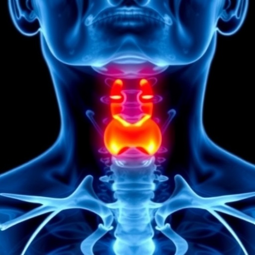
In a groundbreaking advancement for thyroid cancer diagnostics, researchers have developed an innovative radiomics model using nonenhanced computed tomography (NECT) scans to detect papillary thyroid carcinoma (PTC) in patients afflicted with Hashimoto’s thyroiditis (HT). This novel approach addresses the longstanding challenge of identifying PTC amid the diffuse and complex thyroid tissue changes induced by HT—an autoimmune condition that significantly complicates conventional imaging interpretations. The study, published in the prestigious journal BMC Cancer, demonstrates promising improvements in the sensitivity and specificity of PTC detection, potentially transforming early intervention strategies for at-risk patients.
Hashimoto’s thyroiditis represents one of the most common benign thyroid disorders globally, characterized by chronic lymphocytic infiltration and progressive thyroid tissue destruction. Despite its benign classification, HT frequently coexists with PTC, the most prevalent form of thyroid cancer. The coexistence of these two conditions creates substantial diagnostic ambiguity; the inflammatory and fibrotic changes brought on by HT often mask or mimic malignancies on standard imaging modalities such as ultrasound and contrast-enhanced CT scans. These diagnostic difficulties delay treatment and diminish patient outcomes, highlighting the urgent need for more precise diagnostic techniques.
Radiomics—a cutting-edge field leveraging advanced algorithms to extract high-dimensional quantitative features from medical images—has emerged as a powerful tool for oncology diagnostics. By capturing subtle and complex imaging patterns imperceptible to the human eye, radiomics can reveal intrinsic tumor characteristics and microenvironmental heterogeneity. In this study, researchers harnessed the potential of radiomics to analyze NECT images of patients with HT, circumventing the limitations imposed by contrast agents and providing a safer, more accessible diagnostic modality.
The retrospective analysis incorporated data from 130 patients diagnosed pathologically with HT, with or without concurrent PTC. These patients underwent NECT imaging prior to surgical intervention at two distinct medical centers between January 2017 and April 2023. The cohort from Hospital I was partitioned into training and internal validation groups, while data from Hospital II served as an external validation set, ensuring the robustness and generalizability of the model across different clinical settings.
Feature extraction was executed using PyRadiomics, a widely recognized open-source platform facilitating high-throughput quantification of imaging features. Given the complexity of the data—initially comprising hundreds of radiomic features—the research team employed stringent selection criteria. Intraclass correlation coefficients ensured feature reproducibility, Pearson correlation analyses reduced redundant variables, and least absolute shrinkage and selection operator (LASSO) regression identified the most predictive attributes, ultimately condensing the feature set to six pivotal biomarkers.
A critical step involved integrating these refined features into powerful machine learning classifiers to build predictive models. Four algorithms were tested: logistic regression (LR), naive Bayes (NB), support vector machine (SVM), and multilayer perceptron (MLP). This multifaceted approach allowed for comparative evaluation of model performance metrics such as accuracy, sensitivity, specificity, and area under the receiver operating characteristic curve (AUC). The comprehensive comparison underscored the superior performance of the MLP classifier.
In the external validation cohort, the MLP model distinguished itself by achieving an AUC of 0.783, coupled with a sensitivity of 64.3% and a remarkable specificity of 92.3%. These figures indicate the model’s proficient ability to correctly identify true positive cases of PTC while minimizing false positives—a balance crucial for clinical decision-making and avoiding unnecessary invasive procedures. Compared to traditional diagnostic techniques, this radiomics-based model offers a substantial leap in early PTC detection within a challenging clinical population.
The implications of this study are profound. Early and accurate identification of PTC in patients with HT could revolutionize management by facilitating timely surgical and therapeutic interventions, which are pivotal in improving patient prognosis. Furthermore, the use of NECT-based radiomics sidesteps potential adverse reactions linked to contrast agents, broadening its applicability in patients with contraindications for contrast media. This technology also portends significant economic benefits by potentially reducing diagnostic workloads and healthcare expenses associated with misdiagnosis or repeated imaging.
From a technical standpoint, the integration of advanced feature extraction and machine learning exemplifies the transformative impact of artificial intelligence in medical imaging. The researchers’ meticulous methodology, including external validation, enhances confidence in the reproducibility and clinical utility of the model. Moreover, their use of an MLP—a type of artificial neural network adept at capturing nonlinear relationships—reflects a trend toward increasingly sophisticated computational strategies in diagnostic radiology.
This study also signals a paradigm shift toward personalized medicine in thyroid cancer care. By unraveling complex phenotypic patterns hidden within conventional imaging, radiomics can identify patient-specific disease signatures, enabling tailored therapeutic decisions and prognostic assessments. Future research may build upon these findings by incorporating multi-modal imaging data or integrating radiogenomic analyses to further delineate tumor biology and improve predictive accuracy.
While the current model demonstrates considerable prowess, the authors acknowledge limitations including retrospective design, the relatively modest sample size, and potential selection biases inherent in single-country cohorts. They advocate for prospective multicenter trials with larger, more heterogeneous populations to validate and refine the model, ultimately aiming for widespread clinical integration.
In conclusion, the introduction of a NECT-based radiomics model for detecting papillary thyroid carcinoma in Hashimoto’s thyroiditis patients offers a promising leap forward in thyroid oncology diagnostics. By addressing the unique imaging challenges posed by HT, this approach enhances the early detection capabilities, paving the way for improved clinical outcomes. As AI-driven radiomics continues to evolve, its adoption in routine clinical workflows may soon become a critical facet of precision medicine in thyroid disorders and beyond.
Subject of Research: Nonenhanced CT radiomics model development for improved papillary thyroid carcinoma detection in patients with Hashimoto’s thyroiditis.
Article Title: Nonenhanced CT-Based Radiomics Model Enhances PTC Detection in Hashimoto’s Thyroiditis
Article References:
Peng, Y., Huang, K., Gong, Z. et al. Nonenhanced CT-Based radiomics model enhances PTC detection in Hashimoto’s thyroiditis. BMC Cancer 25, 1760 (2025). https://doi.org/10.1186/s12885-025-15206-5
Image Credits: Scienmag.com
DOI: 10.1186/s12885-025-15206-5
Keywords: Radiomics, Nonenhanced CT, Papillary Thyroid Carcinoma, Hashimoto’s Thyroiditis, Machine Learning, Artificial Intelligence, Multilayer Perceptron, LASSO Regression, Medical Imaging, Early Cancer Detection


