


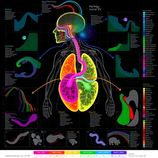
Obesity is often described as a problem of excess body fat, but a new study suggests that its biological footprint is far more extensive—and far more persistent—than changes on the bathroom scale reveal. In a comprehensive analysis of male mice, researchers have built a multi-organ proteomic atlas tracing how obesity reshapes the molecular machinery of the body and how that machinery responds when the animals lose weight. The findings show that recovery is not a single, synchronized process. Instead, different organs appear to follow their own timelines, with some returning rapidly toward a lean state while others retain molecular memories of obesity long after body weight has declined.
Published in Nature Metabolism, the study by Boel, Klinggaard, Akimov and colleagues measured proteins across 15 organs at four stages of diet-induced obesity and regression. In total, the researchers quantified 12,936 unique proteins, creating one of the broadest system-wide maps yet of obesity and weight-loss recovery in an animal model. Proteins are the functional components of cells: they form structural machinery, catalyze chemical reactions, transmit signals, regulate immunity and control the disposal of damaged material. By tracking thousands of these molecules simultaneously, proteomics can reveal biological changes that may not be visible through body weight, blood chemistry or conventional tissue inspection alone.
The scale of the project was made possible by a standardized, semi-automated sample-preparation workflow. Preparing samples from many organs is technically challenging because tissues differ dramatically in their composition. Fat, muscle, brain, blood-rich organs and mineralized tissues each require different handling strategies, and inconsistent preparation can introduce experimental variation that obscures genuine biological signals. By applying a harmonized workflow across the organs, the researchers were able to compare protein abundance across tissues and timepoints with greater consistency. The resulting dataset provides a molecular reference for asking not only which proteins change during obesity, but also whether those changes are shared throughout the body or restricted to particular organs.
The dominant pattern was tissue dependence. Obesity did not produce one universal proteomic signature that appeared identically everywhere. Instead, each organ responded according to its own function, cellular composition and exposure to metabolic stress. Only a relatively small group of proteins changed in the same direction across multiple tissues, suggesting that systemic obesity is assembled from many organ-specific responses rather than driven by a single molecular program. This result may help explain why obesity can produce such a wide range of complications, including insulin resistance, inflammation, altered immune activity, kidney dysfunction and neurological effects. The same metabolic condition can reach different organs through distinct biological routes.
Weight loss restored the proteomes of most tissues toward levels observed in lean animals, indicating that substantial molecular recovery is possible. Yet the return was neither complete nor uniform. White adipose tissue, the main site of energy storage, remained especially altered after regression. The tissue retained strong signatures of phagocytic and inflammatory activity, pointing to continued engagement of immune cells and tissue-remodeling processes. Phagocytosis is the process by which cells engulf and remove debris, dead cells or foreign material. In adipose tissue, persistent phagocytic activity may reflect the cleanup of obesity-associated damage, but it may also indicate that the local immune environment remains activated even after excess weight has been reduced.
The persistence of these signals is important because adipose tissue is not merely a passive storage depot. It acts as an endocrine and immune organ, releasing hormones, cytokines and other signaling molecules that influence the liver, muscle, brain and immune system. If white fat remains inflamed after weight loss, it could continue to affect whole-body physiology even when conventional measures suggest improvement. The researchers used ligand-target inference to investigate the communication networks underlying adipose tissue-specific immune regulation. This computational approach estimates how signaling molecules, or ligands, may influence target proteins or cellular programs in receiving cells. The analysis helped identify candidate regulators that could coordinate the lingering immune state within adipose tissue, offering possible targets for future experimental studies.
The atlas also exposed a separate vulnerability in brown adipose tissue, which is specialized for heat production and energy expenditure. Researchers observed reduced expression of proteasomal subunits in brown fat during obesity. The proteasome is a large protein-degradation complex that breaks down proteins marked for destruction, commonly through attachment of ubiquitin molecules. This ubiquitin–proteasome system is essential for maintaining protein quality, removing faulty or surplus proteins and allowing cells to adapt to changing conditions. When proteasomal components decline, ubiquitin-tagged proteins may not be cleared efficiently. The researchers describe this as stalled ubiquitin turnover, a disruption that could interfere with the performance of brown fat cells and their ability to manage metabolic stress.
The study further indicates that some organs do not show their most prominent obesity-induced changes immediately. The brain, kidney, bone, thymus and spleen displayed delayed alterations, emphasizing that the biological consequences of obesity can unfold on different schedules. Such delayed responses may arise because these organs experience secondary effects of prolonged metabolic imbalance, chronic inflammation or altered hormonal signals rather than direct exposure to excess nutrients alone. The thymus and spleen, central components of immune regulation, may be particularly sensitive to long-term changes in immune-cell development and trafficking. In bone, metabolic and inflammatory signals can influence the activity of cells responsible for building and resorbing tissue. In the brain and kidney, changes may reflect cumulative stress that becomes detectable only after obesity has been established for some time.
These observations challenge the idea that weight loss automatically resets every organ at once. They also suggest that clinical recovery may be better understood as a sequence of partially overlapping processes. A person can experience improvements in blood glucose, blood pressure or liver fat while other tissues remain in a state shaped by earlier metabolic stress. The mouse findings do not establish how long comparable changes persist in humans, nor do they prove that every molecular alteration causes disease. The experiments were conducted in male mice, and sex, age, genetics, diet composition, weight-loss method and duration of obesity could all influence the pace and completeness of recovery. Even so, the atlas offers a valuable framework for testing which molecular changes are temporary adaptations, which represent lingering damage and which may actively prevent full restoration of tissue function.
The researchers have made the datasets openly accessible and developed an interactive webtool intended to support further investigation by the scientific community. Such resources can allow researchers to examine individual proteins, compare organs, follow changes across the four timepoints and generate hypotheses about inter-organ communication. The broader message is that obesity regression is not simply the reversal of weight gain. It is a complex biological transition in which tissues recover, remodel and, in some cases, remain locked in altered states. By documenting those trajectories across the body, the new atlas turns a familiar question—what happens after weight loss?—into a detailed molecular map of recovery, resistance and the possibility of therapeutic intervention.
Multi-organ proteomic changes associated with diet-induced obesity and weight-loss regression in male mice.
Multi-organ proteomic atlas of obesity regression in male mice
Cite Scienmag News
Daisy Hatcher. (August 21, 2026). Proteomic Atlas Maps Obesity Reversal Across Organs in Male Mice. Scienmag. https://scienmag.com/proteomic-atlas-maps-obesity-reversal-across-organs-in-male-mice/
Daisy Hatcher. "Proteomic Atlas Maps Obesity Reversal Across Organs in Male Mice." Scienmag, 21 August 2026, https://scienmag.com/proteomic-atlas-maps-obesity-reversal-across-organs-in-male-mice/. Accessed 4 September 2026.
Daisy Hatcher. "Proteomic Atlas Maps Obesity Reversal Across Organs in Male Mice." Scienmag. August 21, 2026. https://scienmag.com/proteomic-atlas-maps-obesity-reversal-across-organs-in-male-mice/


