


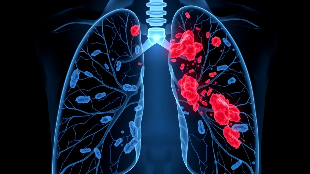
A 71-year-old man who spent years around pigeons arrived at a hospital in Yunnan, China, with a high fever, a stubborn cough, and purulent sputum that gave no hint of what was really driving his illness. Within days he had spiraled from an ordinary-looking chest infection into severe community-acquired pneumonia, type I respiratory failure, and sepsis, the life-threatening body-wide response to infection that pushes blood pressure down and organs toward failure. Routine microbiological tests, the kind most hospitals run first, came back negative, leaving his clinicians to treat a disease they could not name. What followed, documented in a case report published in BMC Infectious Diseases, illustrates both the diagnostic blind spots that still plague severe pneumonia care and the technology that is steadily closing them.
The culprit, when it was finally identified, turned out to be not one pathogen but two. Metagenomic next-generation sequencing, or mNGS, performed on fluid sampled from deep within his lungs detected Chlamydia psittaci, an intracellular bacterium better known as the cause of psittacosis or parrot fever. Meanwhile, conventional sputum culture grew extended-spectrum beta-lactamase-producing Escherichia coli, a multidrug-resistant Gram-negative bacterium whose enzymes can dismantle many of the penicillin- and cephalosporin-class antibiotics that form the backbone of pneumonia therapy. The combination is genuinely rare. C. psittaci pneumonia is uncommon even on its own, typically tied to contact with infected birds, and its coexistence with a drug-resistant enteric bacterium in the same diseased lungs created a therapeutic puzzle that standard treatment guidelines are not built to solve.
The patient’s avian exposure was the first clue that many clinicians might have missed. Long-term contact with pigeons is a recognized risk factor for C. psittaci, which birds carry asymptomatically in their gastrointestinal tracts and shed in droppings, feather dust, and respiratory secretions. Humans inhale the organism in dried aerosols, and the resulting pneumonia can range from a mild flu-like illness to fulminant respiratory failure. Because the bacterium lives inside host cells and grows slowly, standard cultures often fail to detect it, and serological tests require paired samples taken weeks apart, a luxury that a deteriorating patient in an intensive care setting cannot afford. In this case, the initial workup was entirely unrevealing, and the man was begun on empiric therapy with piperacillin-tazobactam, a broad-spectrum beta-lactam and beta-lactamase inhibitor combination, plus doxycycline, the tetracycline-class antibiotic that is the traditional mainstay of psittacosis treatment.
On paper, that regimen should have covered both organisms. Piperacillin-tazobactam retains activity against many ESBL-producing E. coli strains, and doxycycline targets C. psittaci directly. Yet the patient failed to improve, a clinical signal that something in the equation was wrong. It was at this point that the diagnostic team turned to metagenomic next-generation sequencing, a technique that sidesteps the need to culture an organism at all. Instead of trying to grow a specific microbe, mNGS extracts all genetic material from a clinical sample, in this case bronchoalveolar lavage fluid suctioned through a bronchoscope into the deepest airways, and sequences it in bulk. Bioinformatic pipelines then subtract human reads and match the remaining fragments against databases of microbial genomes, producing an unbiased census of everything living in the sample, bacteria, viruses, fungi, and atypical organisms alike.
The sequencing results were decisive. C. psittaci DNA was detected in the lavage fluid, confirming the intracellular pathogen that routine diagnostics had missed, while the parallel sputum culture independently confirmed the ESBL-producing E. coli. The dual findings forced a reassessment of the entire treatment strategy. Antimicrobial susceptibility testing of the cultured E. coli provided the second half of the evidence, showing which antibiotics the resistant strain would and would not respond to. Guided by this combined molecular and phenotypic picture, the clinicians adjusted therapy to a three-drug regimen of piperacillin-tazobactam, doxycycline, and levofloxacin, a fluoroquinolone that adds activity against both atypical organisms and resistant Gram-negative bacteria. Alongside the antimicrobial escalation, the team provided respiratory support and organ protection, the supportive scaffolding that keeps patients alive long enough for antibiotics to work.
The response was what every clinician hopes for but cannot count on in sepsis. The patient improved steadily and was ultimately discharged, a resolution that the authors attribute directly to the treatment adjustment guided by mNGS and susceptibility data. The case thus becomes a textbook argument for molecular diagnostics in severe pneumonia of unknown origin, particularly when a patient fails empiric therapy. Traditional culture-based methods remain indispensable for susceptibility testing, but they are blind to organisms that will not grow on artificial media, and they can take days to yield results that a critically ill patient may not have. mNGS compresses that timeline and broadens the net, catching zoonotic and atypical pathogens that no standard panel would think to test for.
Yet the report also carries a cautionary note about over-reading molecular results, one that reflects a growing tension in the era of ultra-sensitive diagnostics. The authors emphasize that detecting a multidrug-resistant organism in a respiratory sample should be interpreted cautiously, weighing the possibility of colonization against true infection. The human airway and gut harbor complex microbial communities, and E. coli can colonize the respiratory tract of hospitalized patients without causing disease. Distinguishing a bystander from a pathogen requires clinical correlation, meaning the organism’s presence must fit the patient’s illness pattern, inflammatory markers, imaging, and, crucially, the response to targeted therapy. In this case, the treatment failure on the initial regimen and the eventual improvement after adjustment served as the real-world test of whether the ESBL-producing E. coli was a genuine co-pathogen or an innocent passenger, and the clinical trajectory argued for its relevance.
The broader lesson extends beyond this single hospital room. Severe community-acquired pneumonia remains one of the most common reasons for intensive care admission worldwide, and a substantial fraction of cases never receive a confirmed etiological diagnosis, forcing clinicians into empiric choices that can miss atypical organisms or underestimate resistance. Patients with animal exposure, whether birds, livestock, or wildlife, represent a subgroup where the differential diagnosis widens considerably, encompassing zoonoses such as psittacosis, Q fever, and tularemia that routine testing rarely covers. The authors argue that in such patients who fail initial therapy, mixed infections including drug-resistant bacteria must be actively considered rather than assumed away, and that rapid etiological diagnosis through mNGS can meaningfully redirect care. The finding also underscores the quiet global march of ESBL-producing Enterobacterales, which have moved from hospital wards into community settings, meaning that resistance genes once confined to healthcare-associated infections can now appear in a farmer’s lungs.
For the individual patient, the story ended well, a 71-year-old man with respiratory failure and sepsis walking out of the hospital after a diagnostic odyssey that conventional tools could not resolve. For the field, the case adds to a mounting body of evidence that the future of severe pneumonia diagnosis is untargeted, sequencing-based, and paired with disciplined clinical interpretation. The technique is not without limitations, including cost, turnaround variability, contamination risk, and the persistent difficulty of distinguishing colonization from infection, but as this report demonstrates, when a patient with bird exposure deteriorates despite seemingly appropriate antibiotics, the answer may be hiding in two organisms at once, and only an unbiased look at the lung’s microbial census will reveal it.
Subject of Research: A case report of severe community-acquired pneumonia involving co-infection with Chlamydia psittaci and ESBL-producing Escherichia coli diagnosed by metagenomic next-generation sequencing.
Article Title: Severe community-acquired pneumonia complicated by co-infection with Chlamydia psittaci and ESBL-producing Escherichia coli: a case report
Article References: Lu, C., Xie, T., Xu, B., Li, Y., & Yurui, Y. (2026). Severe community-acquired pneumonia complicated by co-infection with Chlamydia psittaci and ESBL-producing Escherichia coli: a case report. BMC Infectious Diseases. https://doi.org/10.1186/s12879-026-14001-2
Image Credits: AI Generated
DOI: 10.1186/s12879-026-14001-2
Keywords: Chlamydia psittaci, psittacosis, community-acquired pneumonia, ESBL-producing Escherichia coli, metagenomic next-generation sequencing, antimicrobial resistance, sepsis, respiratory failure, avian exposure, bronchoalveolar lavage, doxycycline, case report
Cite Scienmag News
Ophelia Keating. (September 20, 2026). Pigeon-Linked Pneumonia Case Reveals Hidden Double Infection Diagnosed by Sequencing. Scienmag. https://scienmag.com/pigeon-linked-pneumonia-case-reveals-hidden-double-infection-diagnosed-by-sequencing/
Ophelia Keating. "Pigeon-Linked Pneumonia Case Reveals Hidden Double Infection Diagnosed by Sequencing." Scienmag, 20 September 2026, https://scienmag.com/pigeon-linked-pneumonia-case-reveals-hidden-double-infection-diagnosed-by-sequencing/. Accessed 5 October 2026.
Ophelia Keating. "Pigeon-Linked Pneumonia Case Reveals Hidden Double Infection Diagnosed by Sequencing." Scienmag. September 20, 2026. https://scienmag.com/pigeon-linked-pneumonia-case-reveals-hidden-double-infection-diagnosed-by-sequencing/


