In the relentless quest to unravel the mechanisms underlying atherosclerosis, a groundbreaking study published in Cell Death Discovery has illuminated the pivotal role of oscillatory shear stress in driving endothelial-to-mesenchymal transition (EndMT), a crucial event accelerating disease progression. The study authored by Li, Xu, Ju, and collaborators presents compelling evidence that mechanical forces in the vascular microenvironment are not merely passive influences but active biochemical signals that transform cellular identity and function in atherosclerotic plaques.
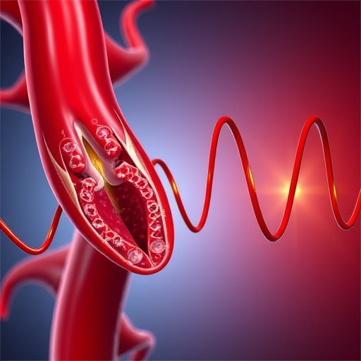
The vascular endothelium, a monolayer of cells lining blood vessels, exists at the interface between circulating blood and the vessel wall. It serves as a dynamic sensor of hemodynamic forces such as shear stress—frictional force generated by blood flow. Under physiological conditions, laminar shear stress promotes endothelial homeostasis, maintaining an anti-inflammatory, anti-thrombotic, and vasodilatory phenotype. However, in regions of disturbed blood flow characterized by oscillatory (bidirectional and low) shear stress, the endothelial cells undergo profound phenotypic changes that predispose these sites to atheroma formation.
This study specifically interrogates how oscillatory shear stress induces EndMT, a process by which endothelial cells lose their characteristic markers and functions while acquiring mesenchymal-like properties, including enhanced migratory capacity, synthesis of extracellular matrix, and a pro-inflammatory profile. EndMT has been increasingly recognized as a pathological mechanism contributing to atherosclerotic plaque progression and instability, yet its mechanical triggering remained poorly understood until now.
Using sophisticated in vitro flow systems mimicking arterial bifurcations where oscillatory shear stress predominates, the authors demonstrated that endothelial cells subjected to this disturbed flow pattern activate an array of mechanotransduction pathways. These include key signaling cascades mediated by transforming growth factor-beta (TGF-β), nuclear factor kappa-light-chain-enhancer of activated B cells (NF-κB), and yes-associated protein (YAP)/transcriptional coactivator with PDZ-binding motif (TAZ), collectively orchestrating the EndMT program. Crucially, the study identified that oscillatory shear stress triggers Smad-dependent TGF-β signaling as a central node integrating mechanical stimuli into nuclear reprogramming, committing endothelial cells towards a mesenchymal fate.
Delving deeper into the molecular intricacies, the research reveals that the mechanosensitive complex formed by platelet endothelial cell adhesion molecule-1 (PECAM-1), vascular endothelial cadherin (VE-cadherin), and vascular endothelial growth factor receptor 2 (VEGFR2) transduces oscillatory shear stress into intracellular biochemical signals. This complex modulates cytoskeletal organization and nuclear mechanotransduction, facilitating chromatin remodeling that enables transcription factors necessary for EndMT to access target gene promoters.
The implications of these findings extend beyond basic biology to clinical relevance. The EndMT-driven fibrotic and inflammatory milieu within plaques enhances extracellular matrix deposition and vulnerability to rupture, which can precipitate myocardial infarction or stroke. Therefore, targeting the biomechanical signaling axis represents a novel therapeutic avenue for stabilizing atherosclerotic lesions. The study’s authors propose that pharmacological modulation of TGF-β signaling or inhibition of YAP/TAZ nuclear translocation could mitigate the EndMT process and its deleterious effects.
Furthermore, the research underscores the heterogeneity of endothelial responses to biomechanical forces, emphasizing that not all shear stress is equal. Protective laminar flows contrast starkly with the pro-pathogenic oscillatory patterns at arterial branches prone to plaque formation. This spatial specificity suggests the potential for localized interventions, such as stent designs engineered to normalize flow patterns or drug delivery systems targeting high-risk vascular regions.
In addition to cellular and molecular experiments, the authors corroborated their in vitro findings by examining human atherosclerotic plaques. Immunohistochemical analyses confirmed the presence of endothelial cells expressing mesenchymal markers within regions exposed to disturbed flow. This in vivo validation strengthens the translational relevance of oscillatory shear stress as a key driver of EndMT in atherosclerosis.
Importantly, this study invites a paradigm shift in our understanding of mechanical forces in vascular biology. Rather than passive contributors to pathology, mechanical cues emerge as dynamic regulators of cellular phenotype and disease trajectory. The integration of biomechanics and molecular signaling offers a fertile ground for interdisciplinary research catalyzing novel diagnostic and therapeutic strategies.
The technological advancements enabling precise simulation of vascular flow patterns and real-time observation of cellular responses underpin the rigor and innovation of this work. High-resolution live-cell imaging combined with transcriptomic profiling allowed the authors to capture the temporal sequence of EndMT induction and trace the signaling pathways activated by direct mechanical stimulation.
Moreover, the findings highlight the complexity of mechanotransduction networks, revealing cross-talk between canonical pathways and novel players that collectively determine endothelial fate. This complexity underscores the challenges and opportunities for targeted interventions, necessitating highly specific and context-dependent therapeutic approaches.
Looking forward, the study paves the way for further exploration into how other biomechanical stimuli—such as cyclic strain or hydrostatic pressure—interact with oscillatory shear stress to influence endothelial behavior and atherogenesis. Additionally, the role of endothelial heterogeneity and the interaction with immune cells in the microenvironment warrant deeper investigation to fully map the landscape of flow-driven vascular pathology.
In sum, this landmark article elucidates how oscillatory shear stress functions as a critical mechanical signal transduction mechanism precipitating endothelial-to-mesenchymal transition and thereby accelerating atherosclerosis progression. The integration of mechanical forces into the molecular framework governing vascular health opens promising horizons for combating cardiovascular disease, a leading cause of morbidity and mortality worldwide.
The insights garnered from this study not only enhance our comprehension of vascular biology but also inspire innovative strategies to diagnose, prevent, and treat atherosclerosis. By manipulating the biomechanical microenvironment or interrupting mechanosensitive signaling pathways, future therapies may halt or even reverse endothelial maladaptation at the earliest stages of disease.
As such, the work by Li, Xu, Ju, and colleagues represents a major advance in cardiovascular research, marrying cutting-edge bioengineering with molecular and cellular pathophysiology. It exemplifies how multidisciplinary approaches can yield transformative understanding and lay the groundwork for next-generation interventions that target the mechanobiology of vascular disease.
Subject of Research: Oscillatory shear stress-induced endothelial-to-mesenchymal transition in atherosclerosis progression
Article Title: Oscillatory shear stress-driven endothelial-to-mesenchymal transition: a critical mechanical signal transduction mechanism in atherosclerosis progression
Article References:
Li, J., Xu, W., Ju, J. et al. Oscillatory shear stress-driven endothelial-to-mesenchymal transition: a critical mechanical signal transduction mechanism in atherosclerosis progression. Cell Death Discov. (2026). https://doi.org/10.1038/s41420-026-03000-6
Image Credits: AI Generated
