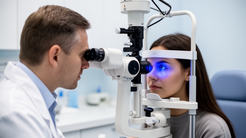
Researchers at the Eye and ENT Hospital of Fudan University in Shanghai have established a stage-specific precision benchmark for measuring corneal epithelial aberrations with the MS-39, a new hybrid device that combines spectral-domain optical coherence tomography with Placido-disk topography. In a prospective study of 141 eyes from 141 patients with keratoconus, the device showed moderate-to-good repeatability and reproducibility across forme fruste, mild, moderate, and total keratoconus, while precision declined noticeably in severe disease. The findings, published in Eye and Vision, provide clinicians with quantitative thresholds for deciding when a measured change reflects true epithelial remodeling rather than random measurement error.
Keratoconus is a degenerative disorder in which the cornea progressively protrudes into a cone shape. Eyes with the condition display a characteristic doughnut-like epithelial thickness profile, with thinning of the epithelium over the cone surrounded by a ring of thicker epithelium. As severity increases, this thinning over the cone becomes more pronounced. The epithelial remodeling masks irregularities of the underlying stromal surface, which makes corneal epithelial aberrations a potentially relevant morphological indicator of the disease. Aberrations describe changes in the optical properties of the eye and can inform diagnosis, treatment, and follow-up, yet the understanding of corneal epithelial aberrations has remained limited.
The MS-39 device acquires corneal epithelial aberrations directly without removing the epithelium, using an 845-nanometer superluminescent light-emitting diode that delivers an axial resolution of 3.6 micrometers in tissue and a transverse resolution of 35 micrometers in air. Each acquisition employs 25 radial scans spanning a 16-millimeter diameter, with 31,232 measured points on the anterior corneal surface and 25,600 on the posterior surface. The interface between the epithelium and Bowman’s layer is detected by a graph search from the OCT B-scans, and epithelial thickness is defined as the vertical distance from the tear film layer to Bowman’s layer. Aberrations are analyzed within a 6-millimeter pupil zone using Zernike decomposition with 36 polynomials and a net refractive index of 1.3375.
The study enrolled patients between January 2021 and August 2023. Keratoconus was diagnosed based on vision loss together with at least one biomicroscopic sign such as focal corneal stromal thinning, Vogt’s striae, or Fleischer ring, combined with abnormal Pentacam topography findings including a maximum keratometry value above 47.2 diopters, an inferior-superior difference above 1.4, or a keratoconus percentage index above 100 percent. Forme fruste keratoconus was defined as the fellow eye of a clinically diagnosed keratoconus eye without abnormal topography or clinical signs. Using the Topographical Keratoconus Classification system, patients were categorized as mild, moderate, or severe while masked to all MS-39 results. Only one eye per subject was included, selected randomly when both eyes qualified.
The final cohort comprised 141 patients, 94 males and 47 females, divided into 32 eyes with forme fruste keratoconus, 29 with mild, 45 with moderate, and 35 with severe disease. Two skilled operators each performed three consecutive measurements on every eye. Measurements were taken in a dark room without pupil dilation, between 10 a.m. and 5 p.m. to minimize daily corneal variation, and topical eye drops were prohibited for two hours beforehand. Only acquisitions meeting strict quality criteria, including at least 90 percent image coverage, were analyzed. The evaluated parameters were total root-mean-square aberrations, higher-order aberration RMS, coma, trefoil, spherical aberration, and astigmatism II.
The precision results showed that in the forme fruste, mild, moderate, and total groups, the within-subject standard deviation for intraobserver repeatability was at most 0.18 micrometers and the test-retest variability at most 0.50 micrometers. Intraclass correlation coefficients reached at least 0.884 for most parameters, with the exceptions of trefoil, ranging from 0.493 to 0.797, and astigmatism II, ranging from 0.548 to 0.859. For interobserver reproducibility, the corresponding values were even tighter, with within-subject standard deviations at most 0.15 micrometers, test-retest variability at most 0.41 micrometers, and intraclass correlation coefficients of at least 0.917 except for trefoil and astigmatism II.
Severe keratoconus told a different story. In that subgroup, intraclass correlation coefficients for intraobserver repeatability ranged from 0.366 to 0.904, and for interobserver reproducibility from 0.493 to 0.908. Within-subject standard deviations rose to between 0.13 and 0.28 micrometers for repeatability and 0.15 to 0.25 micrometers for reproducibility, with test-retest values spanning 0.36 to 0.77 micrometers and 0.42 to 0.69 micrometers respectively. Bland-Altman analysis found no clinically significant systematic bias between operators, with mean differences below 0.1 micrometers and 95 percent confidence intervals crossing zero, indicating that the reduced reliability stemmed from increased random measurement error rather than operator-related bias. The authors attribute this to marked corneal irregularity, epithelial thinning that challenges edge detection, and tear film instability in advanced disease.
The researchers also emphasized an important statistical caveat when interpreting intraclass correlation coefficients. The ICC reflects the proportion of total variance attributable to true intersubject biological differences and is therefore influenced by both random measurement error and the variability within the study sample. For parameters with inherently low numerical dispersion, such as trefoil and astigmatism II in early keratoconus, even minimal absolute measurement error can produce a moderate or low ICC that does not necessarily indicate clinically insufficient precision. For this reason, the team prioritized test-retest variability as the primary endpoint, since it represents the minimal detectable change at the 95 percent confidence level.
Building on these results, the authors proposed stage-specific test-retest thresholds for clinical interpretation. For forme fruste, mild, moderate, and total keratoconus, all parameters met a uniform threshold of 0.50 micrometers or less, meaning changes exceeding that value can be reliably interpreted as biological epithelial remodeling rather than measurement noise. More granular thresholds for total RMS, HOA RMS, coma, trefoil, spherical aberration, and astigmatism II were 0.31, 0.24, 0.20, 0.20, 0.19, and 0.21 micrometers for forme fruste keratoconus; 0.33, 0.25, 0.25, 0.28, 0.21, and 0.32 micrometers for mild keratoconus; and 0.50, 0.41, 0.32, 0.30, 0.31, and 0.35 micrometers for moderate keratoconus. In severe disease, the authors advise extreme caution and do not recommend the device as the sole basis for clinical decisions.
The study had limitations. No other principle-based devices were included for comparison, and device-specific differences in epithelial and Bowman’s layer metrics across OCT platforms may affect clinical application. Objective tear film metrics were not quantitatively assessed, although strict image quality criteria were applied to exclude unstable conditions. The authors plan future work involving patients with more severe keratoconus and other corneal ectatic diseases such as pellucid marginal degeneration. As the first study to systematically evaluate MS-39 epithelial aberration precision stratified by disease severity, the work offers a practical reference for clinicians tracking keratoconus progression, supporting the device’s potential for early diagnosis and longitudinal monitoring while underscoring that precision must be tailored to disease stage.
Subject of Research: Measurement precision of corneal epithelial aberrations across keratoconus stages using a hybrid SD-OCT and Placido-disk device
Article Title: Establishing a precision reference benchmark for corneal epithelial aberration measurements using a new hybrid device across keratoconus stages
Article References: Yang, Y., Ning, R., Li, S., Yang, X., Wang, Y., Li, K., Savini, G., Schiano-Lomoriello, D., Zhou, X., & Huang, J. (2026). Establishing a precision reference benchmark for corneal epithelial aberration measurements using a new hybrid device across keratoconus stages. Eye and Vision, 13(1), Article 38. https://doi.org/10.1186/s40662-026-00508-x
Image Credits: AI Generated
DOI: 10.1186/s40662-026-00508-x
Keywords: keratoconus, corneal epithelial aberrations, MS-39, optical coherence tomography, Placido-disk topography, repeatability, reproducibility, test-retest variability, forme fruste keratoconus, ophthalmic imaging, Bowman's layer, Zernike analysis
Cite Scienmag News
Ophelia Keating. (October 29, 2026). New Hybrid Imaging Device Sets Precision Benchmark for Keratoconus Stages. Scienmag. https://scienmag.com/new-hybrid-imaging-device-sets-precision-benchmark-for-keratoconus-stages/
Ophelia Keating. "New Hybrid Imaging Device Sets Precision Benchmark for Keratoconus Stages." Scienmag, 29 October 2026, https://scienmag.com/new-hybrid-imaging-device-sets-precision-benchmark-for-keratoconus-stages/. Accessed 30 October 2026.
Ophelia Keating. "New Hybrid Imaging Device Sets Precision Benchmark for Keratoconus Stages." Scienmag. October 29, 2026. https://scienmag.com/new-hybrid-imaging-device-sets-precision-benchmark-for-keratoconus-stages/