


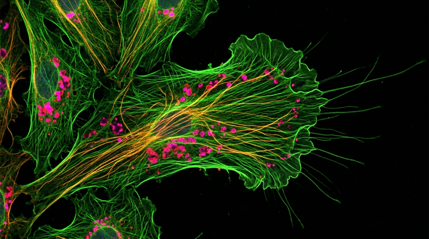
Researchers have developed a multiplexed confocal fluorescence lifetime imaging microscopy (FLIM) system capable of capturing dynamic molecular processes in live cells at a frame rate of 4 Hz. Published in the journal Discover Imaging in May 2025, the study presents a 32 by 32 point multiplexing time-resolved confocal microscope designed to overcome the speed limitations of conventional single-point confocal systems. The primary objective of this engineering advancement is to enable quantitative measurements of biomolecular interactions with high spatial, spectral, and temporal resolution, addressing a critical bottleneck in biological imaging where phototoxicity and photobleaching often restrict excitation power in live specimens.
The core challenge in live-cell FLIM has historically been the trade-off between image acquisition speed and signal fidelity. Conventional confocal microscopy acquires images one pixel at a time, a process that is inherently slow and requires high laser power to gather sufficient photons within short dwell times. This high power can damage live cells, while low power results in poor signal-to-noise ratios. The new system utilizes a pinhole array to simplify the optical design, improving optical efficiency and allowing for faster frame rates without compromising the confocal resolution necessary for subcellular imaging.
The optical architecture of the new microscope relies on a pulsed diode laser emitting 440 nm pulses with a repetition rate of 50 MHz. The laser beam is coupled through a multimode fiber to ensure uniformity before being collimated into a 1.4 cm beam. A 32 by 32 microlens array (MLA) generates 1024 points of excitation, enabling parallel acquisition of multiple pixels. This multiplexing approach significantly increases the photon count rate compared to single-point systems, allowing the use of laser powers compatible with live-cell imaging while maintaining high contrast in complex environments.
Central to the system’s high-speed capability is the use of a single photon avalanche detector (SPAD) array, specifically the SPC3 model from Micro Photon Devices. This detector is capable of measuring three distinct temporal gates, which are essential for calculating fluorescence lifetimes. The researchers calibrated these temporal gates using a 1 µM solution of Coumarin6, a fluorophore with a known mono-exponential decay. By adjusting the delay between laser triggering and the start of the first gate, they ensured that the three gates were correctly positioned within the 20 ns laser period, with each gate having a width of 3 ns.
To achieve the target frame rate, the team redesigned the optomechanical scanning system. Previous scanner controls stepped to each pixel position and stopped, introducing delays. The new control system employs a continuous raster scan motion without stopping, moving from the start of imaging to the end in a single fluid motion. Detailed calibration of the galvanometer window scanners revealed that the highest reliable mechanical frame rate is approximately 4.1 frames per second at a 200 µs pixel dwell time. However, to account for data transfer and reset periods, the system operates at a real-time frequency of 3.7 Hz for live-cell imaging, using a 250 µs pixel dwell time.
The study demonstrates the system’s precision through dilution series measurements of Coumarin6 and Fluorescein. At a 250 µs pixel dwell time, the system accurately measured Coumarin6 concentrations as low as 10 µM at 4 Hz frame rates. The researchers established a photon count threshold of 25 photons to ensure accurate lifetime estimation using the rapid lifetime determination (RLD) algorithm. Below this threshold, the signal-to-noise ratio drops, leading to increased errors in lifetime estimation. The use of the SPAD array, with its low noise floor, allows for precise measurement of mono-exponential decays even at these lower concentrations.
Live-cell experiments were conducted using HEK293 T cells transfected with fluorescent proteins mCerulean3 and Venus. The system successfully imaged these cells at high frame rates, revealing subcellular lifetime variations. In regions with different expression ratios of the two proteins, lifetimes were measured at 3.7 ± 0.2 ns and 3.3 ± 0.2 ns with a 1000 µs dwell time, and 3.8 ± 0.3 ns and 3.5 ± 0.3 ns with a 250 µs dwell time. The broader distribution in the shorter exposure dataset was attributed to higher estimation errors in low photon count regions, a predictable consequence of the reduced exposure time.
A key application of this technology is the measurement of Förster Resonance Energy Transfer (FRET), a technique that allows for the indirect observation of molecular interactions. FRET occurs when energy is transferred non-radiatively from an excited donor fluorophore to an acceptor fluorophore, reducing the donor’s lifetime. This process requires the fluorophores to be within approximately 10 nm of each other, making it a sensitive probe for protein binding and conformational changes. The new system demonstrated its capability by measuring FRET in live cells using a construct where mCerulean3 and Venus are linked. The measured lifetime for the FRET construct was 2.3 ± 0.3 ns, significantly lower than the single protein lifetimes, confirming the occurrence of energy transfer.
The system’s performance was further validated by imaging fixed, stained samples, including Convallaria rhizomes stained with Safranin and Fast-Green. The confocal FLIM images revealed lifetime variations around 2.4 ns and 2.9 ns, corresponding to the different binding environments of the stains. These measurements agreed with previous literature values, demonstrating the system’s accuracy in complex, spatially heterogeneous samples. The ability to maintain a full field of view of 960 by 960 pixels at 4 Hz is a substantial improvement over traditional fast confocal FLIM microscopes, which often limit the field of view to 256 by 256 pixels to achieve similar frame rates.
The researchers note that the current frame rate is limited by the mechanical scanning speed of the window scanners rather than the optical design. The improved optical throughput from the pinhole array allows for higher photon count rates, theoretically supporting faster imaging. Future improvements could involve the use of resonant galvanometric scanners, which might allow the technology to reach tens of Hz. Additionally, the current signal processing uses a two-gate RLD method, which computes the average lifetime but does not separate multi-exponential decays. The authors suggest that replacing the gated SPAD array with a time-correlated single photon counting (TCSPC) detector could provide higher temporal resolution and enable the analysis of more complex decay profiles.
This advancement in multiplexed confocal FLIM offers a significant tool for biologists studying dynamic cellular processes. By providing high-speed, high-resolution lifetime imaging, the system allows for the observation of molecular interactions on sub-second timescales, which are critical for understanding signaling pathways and drug mechanisms. The ability to image live cells with minimal photodamage and high temporal resolution opens new possibilities for pharmacological research and the development of personalized treatments. The study highlights the potential of combining advanced detector technology with optimized optical and mechanical designs to push the boundaries of fluorescence lifetime imaging microscopy.
The work was conducted by a team of researchers including Morgan Richards, Nikolina Malic, Elizabeth J. Osterlund, Andrea Rhikkaella Buendia, Laura Polga, Ray Truant, and Qiyin Fang. The paper, titled “Multiplexed confocal FLIM for dynamic molecular imaging in live cells,” was published in Discover Imaging, Volume 2, article number 7, in 2025. The data underlying the results are not publicly available but may be obtained from the authors upon reasonable request. This research contributes to the growing field of high-speed fluorescence imaging, providing a robust platform for investigating the dynamic nature of biological systems at the molecular level.
Subject of Research: Multiplexed confocal FLIM for dynamic molecular imaging in live cells
Article Title: Multiplexed confocal FLIM for dynamic molecular imaging in live cells
Article References: Richards, M., Malic, N., Osterlund, E. J., Buendia, A. R., Polga, L., Truant, R., & Fang, Q. (2025). Multiplexed confocal FLIM for dynamic molecular imaging in live cells. Discover Imaging, 2(1), Article 7. https://doi.org/10.1007/s44352-025-00010-5
Image Credits: AI Generated
DOI: 10.1007/s44352-025-00010-5
Keywords: Multiplexed, confocal, FLIM, dynamic, molecular, imaging, live, cells, scientific research
Cite Scienmag News
Denise Maddox. (November 11, 2026). New Confocal Microscope Captures Live Cell Dynamics at 4 Hz. Scienmag. https://scienmag.com/new-confocal-microscope-captures-live-cell-dynamics-at-4-hz/
Denise Maddox. "New Confocal Microscope Captures Live Cell Dynamics at 4 Hz." Scienmag, 11 November 2026, https://scienmag.com/new-confocal-microscope-captures-live-cell-dynamics-at-4-hz/. Accessed 22 November 2026.
Denise Maddox. "New Confocal Microscope Captures Live Cell Dynamics at 4 Hz." Scienmag. November 11, 2026. https://scienmag.com/new-confocal-microscope-captures-live-cell-dynamics-at-4-hz/




