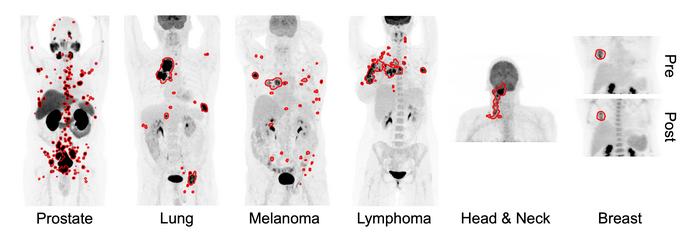
Toronto, Ontario—A novel AI approach can accurately detect six different types of cancer on whole-body PET/CT scans, according to research presented at the 2024 Society of Nuclear Medicine and Molecular Imaging Annual Meeting. By automatically quantifying tumor burden, the new tool can be useful for assessing patient risk, predicting treatment response, and estimating survival.
“Automatic detection and characterization of cancer are important clinical needs to enable early treatment,” said Kevin H. Leung, PhD, research associate at Johns Hopkins University School of Medicine in Baltimore, Maryland. “Most AI models that aim to detect cancer are built on small to moderately sized datasets that usually encompass a single malignancy and/or radiotracer. This represents a critical bottleneck in the current training and evaluation paradigm for AI applications in medical imaging and radiology.”
To address this issue, researchers developed a deep transfer learning approach (a type of AI) for fully automated, whole-body tumor segmentation and prognosis on PET/CT scans. Data from 611 FDG PET/CT scans of patients with lung cancer, melanoma, lymphoma, head and neck cancer, and breast cancer, as well as 408 PSMA PET/CT scans of prostate cancer patients were analyzed in the study.
The AI approach automatically extracted radiomic features and whole-body imaging measures from the predicted tumor segmentations to quantify molecular tumor burden and uptake across all cancer types. Quantitative features and imaging measures were used to build predictive models to demonstrate prognostic value for risk stratification, survival estimation, and prediction of treatment response in patients with cancer.
“In addition to performing cancer prognosis, the approach provides a framework that will help improve patient outcomes and survival by identifying robust predictive biomarkers, characterizing tumor subtypes, and enabling the early detection and treatment of cancer,” noted Leung. “The approach may also assist in the early management of patients with advanced, end-stage disease by identifying appropriate treatment regimens and predicting response to therapies, such as radiopharmaceutical therapy.”
Leung noted that in the future generalizable, fully automated AI tools will play a major role in imaging centers by assisting physicians in interpreting PET/CT scans of patients with cancer. The deep learning approach may also lead to the discovery of important molecular insights about the underlying biological processes that may be currently understudied in large-scale patient populations.
Abstract 241979. “Fully Automated Whole-Body Tumor Segmentation on PET/CT using Deep Transfer Learning,” Kevin Leung, Steven Rowe, Moe Sadaghiani, Jeffrey Leal, Esther Mena, Peter Choyke, Yong Du, Martin Pomper, Johns Hopkins University School of Medicine, Baltimore, Maryland.
###
All 2024 SNMMI Annual Meeting abstracts can be found online.
About the Society of Nuclear Medicine and Molecular Imaging
The Society of Nuclear Medicine and Molecular Imaging (SNMMI) is an international scientific and medical organization dedicated to advancing nuclear medicine and molecular imaging—vital elements of precision medicine that allow diagnosis and treatment to be tailored to individual patients in order to achieve the best possible outcomes.
SNMMI’s members set the standard for molecular imaging and nuclear medicine practice by creating guidelines, sharing information through journals and meetings and leading advocacy on key issues that affect molecular imaging and therapy research and practice. For more information, visit www.snmmi.org.

Credit: Image created by Kevin H. Leung et al., Johns Hopkins University, Baltimore, MD.
Toronto, Ontario—A novel AI approach can accurately detect six different types of cancer on whole-body PET/CT scans, according to research presented at the 2024 Society of Nuclear Medicine and Molecular Imaging Annual Meeting. By automatically quantifying tumor burden, the new tool can be useful for assessing patient risk, predicting treatment response, and estimating survival.
“Automatic detection and characterization of cancer are important clinical needs to enable early treatment,” said Kevin H. Leung, PhD, research associate at Johns Hopkins University School of Medicine in Baltimore, Maryland. “Most AI models that aim to detect cancer are built on small to moderately sized datasets that usually encompass a single malignancy and/or radiotracer. This represents a critical bottleneck in the current training and evaluation paradigm for AI applications in medical imaging and radiology.”
To address this issue, researchers developed a deep transfer learning approach (a type of AI) for fully automated, whole-body tumor segmentation and prognosis on PET/CT scans. Data from 611 FDG PET/CT scans of patients with lung cancer, melanoma, lymphoma, head and neck cancer, and breast cancer, as well as 408 PSMA PET/CT scans of prostate cancer patients were analyzed in the study.
The AI approach automatically extracted radiomic features and whole-body imaging measures from the predicted tumor segmentations to quantify molecular tumor burden and uptake across all cancer types. Quantitative features and imaging measures were used to build predictive models to demonstrate prognostic value for risk stratification, survival estimation, and prediction of treatment response in patients with cancer.
“In addition to performing cancer prognosis, the approach provides a framework that will help improve patient outcomes and survival by identifying robust predictive biomarkers, characterizing tumor subtypes, and enabling the early detection and treatment of cancer,” noted Leung. “The approach may also assist in the early management of patients with advanced, end-stage disease by identifying appropriate treatment regimens and predicting response to therapies, such as radiopharmaceutical therapy.”
Leung noted that in the future generalizable, fully automated AI tools will play a major role in imaging centers by assisting physicians in interpreting PET/CT scans of patients with cancer. The deep learning approach may also lead to the discovery of important molecular insights about the underlying biological processes that may be currently understudied in large-scale patient populations.
Abstract 241979. “Fully Automated Whole-Body Tumor Segmentation on PET/CT using Deep Transfer Learning,” Kevin Leung, Steven Rowe, Moe Sadaghiani, Jeffrey Leal, Esther Mena, Peter Choyke, Yong Du, Martin Pomper, Johns Hopkins University School of Medicine, Baltimore, Maryland.
###
All 2024 SNMMI Annual Meeting abstracts can be found online.
About the Society of Nuclear Medicine and Molecular Imaging
The Society of Nuclear Medicine and Molecular Imaging (SNMMI) is an international scientific and medical organization dedicated to advancing nuclear medicine and molecular imaging—vital elements of precision medicine that allow diagnosis and treatment to be tailored to individual patients in order to achieve the best possible outcomes.
SNMMI’s members set the standard for molecular imaging and nuclear medicine practice by creating guidelines, sharing information through journals and meetings and leading advocacy on key issues that affect molecular imaging and therapy research and practice. For more information, visit www.snmmi.org.
Journal
Journal of Nuclear Medicine
Article Title
Fully Automated Whole-Body Tumor Segmentation on PET/CT using Deep Transfer Learning
Article Publication Date
10-Jun-2024


