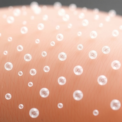
In the microscopic realm of environmental pollutants, nanoplastics have emerged as a silent, pervasive threat that extends far beyond the oceans into previously unconsidered pathways, such as human skin. Dr. Wei Xu and his research team at Texas A&M University have pioneered groundbreaking investigations revealing how nanoplastics, especially those exposed to seawater, interact with and infiltrate skin cells, raising critical questions about their implications for human health. These microscopic particles, often less than 100 nanometers in size, are ubiquitous fragments from the degradation of conventional plastics. Their small size allows them to traverse biological barriers and potentially cause cellular dysfunction, yet their interactions with living tissues remain poorly understood.
To understand the complexity of nanoplastic toxicity, Dr. Xu’s team homed in on the environmental transformations these particles undergo once they enter marine ecosystems. Nanoplastics rapidly acquire a coating of biomolecules such as proteins, organic compounds, and other chemical residues from seawater, effectively creating a “protein corona” that modifies the biological identity and cellular interactions of the particles. This protein corona can shield nanoplastics from recognition by the immune system, altering their fate once inside the human body. Such modifications have profound implications for toxicology, as the environmental history of a particle may dictate its effects on human cells, rather than the particle alone.
The researchers simulated real-world conditions by incubating commercially available nanoplastic beads with seawater collected off the coast of Corpus Christi. This procedure allowed natural marine proteins and compounds to adsorb to the particles, mimicking authentic environmental exposure. Upon subsequent exposure to cultured human skin cells, specifically keratinocytes and fibroblasts, these coated nanoplastics exhibited an unexpected ability to evade cellular degradation pathways. Unlike pristine nanoplastics, which are often recognized and destroyed by lysosomes—the cellular “recycling centers”—the protein-coated particles persisted inside cells, suggesting a form of biological camouflage.
Such persistence raises critical questions regarding the long-term cellular and systemic consequences of nanoplastic accumulation. The research implies that nanoplastics can bypass the cellular “garbage disposal,” potentially leading to intracellular buildup that might disrupt cell function or induce chronic inflammation. More troubling is the possibility that these environmental coatings themselves could harbor toxins or facilitate the transport of harmful substances into the skin, amplifying the biological hazards posed by nanoplastic exposure.
Further, this research extends our understanding of skin as not merely a passive barrier but an active interface susceptible to nanoparticle infiltration. Although ingestion of nanoplastics—through food and water—has received considerable attention, dermal absorption represents an underexplored route for human exposure that could have different physiological outcomes. Dr. Xu’s findings emphasize the need for a more holistic approach to nanoplastic toxicology, integrating environmental context with biological impact.
However, the challenge of characterizing nanoplastic interactions with skin cells is made complex by the variability of environmental coatings. While proteins comprise a major component of the corona studied by Xu’s team, other biological and chemical constituents common in seawater, such as toxins from algal blooms or chemical pollutants from anthropogenic sources, might likewise alter particle behavior. Moreover, environmental fluctuations caused by climate change, such as increased flooding or changes in marine ecosystems, could produce novel coating profiles, requiring continual adaptation of research methodologies and risk assessments.
Standardization of research protocols emerges as a pivotal goal in advancing nanoplastic toxicology. Currently, studies often produce divergent and contradictory results, partly because environmental coatings on nanoplastics are not consistently accounted for. Developing a consensus on how to replicate and include environmental factors in nanoplastic exposure studies will ensure data comparability and improve the reliability of risk evaluations. Xu urges the scientific community to adopt unified approaches in the preparation and testing of nanoplastics to better understand their widespread impact.
Beyond method standardization, the scope of research must expand to systematically identify and characterize the various types of nanoplastic coatings found in diverse environments. The initial discovery of protein coronas on ocean-aged nanoplastics represents only the beginning. Comprehensive profiling of these coatings, including lipids, polysaccharides, and synthetic chemicals, is crucial to formulating a complete picture of nanoplastic interactions with biological systems. Such detailed investigations will provide insights into the molecular mechanisms underlying cellular uptake, immune evasion, and toxicity.
The implications of this work reach far beyond environmental health, touching on public safety, regulatory frameworks, and materials science innovation. Understanding how nanoplastics interact with human cells could inform the development of new materials and coatings designed to minimize environmental persistence or biological absorption. Moreover, elucidating the pathways and effects of nanoplastic exposure is essential for crafting informed policies aimed at reducing plastic pollution and safeguarding human health.
As society grapples with the growing crisis of plastic pollution, Dr. Xu’s research marks a significant advancement in unveiling the subtle yet consequential ways nanoplastics affect human biology. The environmental protein corona concept illustrates that the risks posed by nanoplastics are dynamic and context-dependent, necessitating interdisciplinary efforts crossing environmental science, toxicology, and biomedicine. Through continued exploration, researchers hope to devise innovative interventions to mitigate nanoplastic harm while advancing our comprehension of this emergent pollutant class.
In conclusion, the work spearheaded by Dr. Wei Xu and his colleagues illuminates an urgent, previously underestimated vector of nanoplastic exposure through the skin, demonstrating that environmental transformations critically influence particle behavior and toxicity. This paradigm shift in understanding demands new models of health risk assessment, heightened awareness regarding dermal exposure pathways, and sustained scientific inquiry into the evolving nature of nano-sized pollutants in a world increasingly dominated by plastic waste.
Subject of Research: Cells
Article Title: Environmental protein corona on nanoplastics altered the responses of skin keratinocytes and fibroblast cells to the particles
News Publication Date: 15-Aug-2025
Web References: DOI: 10.1016/j.jhazmat.2025.138722
References: Xu, W. et al. (2025). Environmental protein corona on nanoplastics altered the responses of skin keratinocytes and fibroblast cells to the particles. Journal of Hazardous Materials.
Image Credits: Texas A&M University
Keywords: Nanoparticles, Human health


