In a groundbreaking study published in Translational Psychiatry, researchers have harnessed the power of multimodal cortical parcellations to unveil previously uncharted regions within the human cerebral cortex that bear significant correlations to cognitive performance. This remarkable advancement not only deepens our understanding of the functional architecture of the brain but also paves the way for novel diagnostic and therapeutic strategies aligned with the unique neural landscapes of individuals. As the quest to decipher the enigmatic human brain intensifies, this study emerges as a beacon, demonstrating the potency of integrating diverse imaging modalities to map the cerebral cortex with unprecedented precision.
The cerebral cortex, a thin but intricately folded layer of neural tissue covering the brain’s surface, is the command center for cognition, perception, and voluntary behavior. Traditional neuroscientific approaches have long sought to segment this complex tissue into distinct regions or “parcellations” based on structural and functional features. However, earlier methods often relied on a single imaging modality such as magnetic resonance imaging (MRI), which, while powerful, limits the granularity and functional relevance of the identified regions. The innovation driving this latest work lies in the adoption of multimodal techniques that synthesize different imaging data sources—such as functional MRI (fMRI), diffusion tensor imaging (DTI), and structural MRI—offering a multidimensional view of cortical organization.
Delving deeper into the methodology, the research team implemented a sophisticated framework that leverages complementary data streams to define cortical parcels with greater anatomical fidelity and cognitive significance. Functional MRI provides dynamic insights by measuring blood oxygenation changes reflective of neural activity, capturing how different brain areas engage during cognitive tasks. Diffusion tensor imaging maps white matter tracts, illuminating structural connectivity patterns that underpin inter-regional communication. By combining these modalities with high-resolution anatomical scans, the investigators achieved a comprehensive cortical atlas that transcends mere anatomical landmarks and incorporates functional relevance, connectivity profiles, and microstructural characteristics.
One of the most transformative aspects of this study is the identification of novel cortical subdivisions that had eluded detection through unimodal analyses. These newly discovered areas exhibit distinct patterns of connectivity and activation, suggesting specific roles in cognitive processes such as working memory, attention regulation, and executive control. The implications of uncovering these regions are profound: they offer new targets for understanding the neural substrates of intelligence and cognitive variability in both healthy individuals and neuropsychiatric disorders. Moreover, these insights may help explain why some people excel in certain cognitive domains, opening doors for personalized cognitive enhancement strategies.
Further, the research underscores the dynamic interplay between cortical structure and function, challenging the traditional static view of neuroanatomical divisions. By integrating multimodal data, the scientists demonstrated that cognitive performance is linked not only to the presence of certain cortical areas but to their connectivity profiles and activity patterns under varying cognitive loads. This approach represents a paradigm shift in cognitive neuroscience, emphasizing an integrative perspective that captures the brain’s complexity and its adaptive capacity to support diverse mental operations.
From a clinical standpoint, the discoveries reported in this work promise to revolutionize the diagnosis and treatment of cognitive impairments. Disorders such as schizophrenia, Alzheimer’s disease, and autism spectrum disorders often involve subtle disruptions in cortical organization and connectivity. By providing a refined map of functionally significant cortical parcels, this research enables the identification of atypical patterns that may underlie these conditions. Furthermore, these findings set the stage for the development of biomarker-based approaches, employing neuroimaging data to predict disease risk, monitor progression, and tailor interventions to individual cortical profiles.
The research also offers vital insights into neurodevelopmental trajectories, as parcellation patterns evolve from infancy to adulthood. Understanding how these cortical regions emerge and specialize throughout development sheds light on critical windows for cognitive maturation and the potential impact of environmental and genetic factors. Longitudinal studies expanding on this multimodal parcellation framework could illuminate mechanisms of neuroplasticity and resilience, informing educational strategies and early interventions.
Technological advances undoubtedly played a crucial role in enabling this research. High-field MRI scanners, machine learning algorithms for image analysis, and advanced data fusion methods collectively facilitated the extraction and integration of complex brain features. The study’s success highlights the increasingly interdisciplinary nature of neuroscience, where computational science, engineering, and biology converge to tackle some of the most intricate challenges.
Importantly, the study’s findings provoke broader questions about the very definition of brain regions. Traditional brain atlases, while useful, often suffer from inconsistencies and lack sensitivity to individual differences. The novel multimodal parcellations provide a more personalized and nuanced brain map, potentially redefining neuroanatomical nomenclature and guiding future research toward individualized neuroscience—a frontier aligned with precision medicine.
As the field moves forward, the data from this study offer a valuable resource for researchers aiming to connect genotype, brain phenotype, and cognitive behavior. Integrating cortical parcellation maps with genetic, epigenetic, and environmental data sets may unlock complex mechanisms governing cognition and brain health. Such integrative approaches could ultimately lead to breakthroughs in enhancing cognitive capacities and mitigating deficits across the lifespan.
Moreover, the study emphasizes the importance of open science and data sharing. By making their cortical parcellation maps and analytical pipelines accessible to the scientific community, the authors foster collaborative efforts that accelerate discoveries. This cooperative model ensures that the insights and tools generated extend beyond a single study, catalyzing a broader transformation in neuroscience research methodologies.
The implications of these findings are not confined solely to academic circles but extend into educational domains and public health policy. By elucidating specific brain regions linked to cognitive strengths and weaknesses, educators and clinicians can design targeted training programs to maximize cognitive potential or rehabilitate impaired functions. Policymakers might utilize such scientific evidence to allocate resources toward mental health initiatives that are informed by cutting-edge neuroscience.
Finally, this study acts as a clarion call to revisit how we conceptualize and study the brain in both health and disease. It encourages a move away from reductionist models toward embracing the brain’s multifaceted nature as revealed through integrative multimodal imaging. As neuroscience continues to evolve, studies like this illuminate pathways to unlock the mysteries of cognitive function, transforming our understanding of the human mind and its boundless capabilities.
In summary, the deployment of multimodal cortical parcellations marks a transformative step in cognitive neuroscience, providing unprecedented clarity into the cerebral cortex’s organization and its relationship with cognitive performance. This innovative approach not only identifies novel functionally significant brain regions but also redefines how we explore and interpret individual variability in cognition. The translational potential of these findings heralds a future where precision maps of the brain inform diagnosis, treatment, and enhancement of cognitive function, offering hope and insight into the intricacies of the human mind.
Subject of Research: Identification of novel cerebral cortex regions related to cognitive performance using multimodal cortical parcellations.
Article Title: Using multimodal cortical parcellations to identify novel regions of the human cerebral cortex associated with cognitive performance.
Article References:
Qiu, S., Zhang, Z., Liang, H. et al. Using multimodal cortical parcellations to identify novel regions of the human cerebral cortex associated with cognitive performance. Transl Psychiatry (2026). https://doi.org/10.1038/s41398-025-03803-8
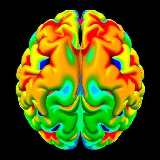
Image Credits: AI Generated
