


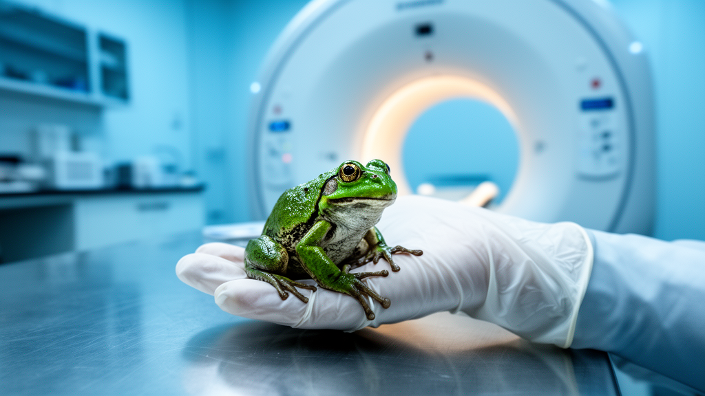
Telling a boy frog from a girl frog sounds simple, but for veterinarians and conservationists working with young amphibians it is one of the field’s most stubborn problems. A team of Hungarian researchers has now put three standard imaging tools—ultrasound, computed tomography, and magnetic resonance imaging—to a formal test in small, immature agile frogs, and the results reveal both a surprising success and a sobering limit. MRI could pick out testes in juvenile males that no other scanner could see, yet immature ovaries slipped past every machine. The study, published in Frontiers in Veterinary Science, offers the first systematic validation of minimally invasive sexing techniques in young frogs, a gap that has long frustrated breeding programs racing to save endangered amphibians.
The stakes are higher than they might appear. More than 2,000 amphibian species are listed as endangered, and populations are declining worldwide, making captive breeding and reintroduction programs critical lifelines. Knowing the sex of individual animals is essential for pairing breeders, managing genetic diversity, and timing reintroductions. Yet many amphibians show no external signs of sex until maturity, and even adults can deceive. Body size overlaps substantially between the sexes, offering only about 70 to 75 percent accuracy in adult frogs. Hormone measurements from blood, urine, or feces can be thrown off by seasonal cycles, stress, or captive housing conditions. Nuptial pads on males’ thumbs and other secondary traits often appear only around the breeding season, and the absence of environmental cues in captivity can suppress them entirely.
Genetic testing might seem like the obvious answer, but amphibian genetics complicates rather than simplifies matters. Most amphibians lack distinct sex chromosomes of the kind that make molecular sexing routine in mammals and birds, and polyploidy is common, so researchers must develop species-specific genetic markers before a single swab can be read. Worse, genotypic sex can be overridden by environmental influences during embryonic or larval development, meaning an animal’s chromosomes and its actual, functional sex may not match. For conservation programs that need to know which animals will lay eggs and which will fertilize them, phenotypic sex—the sex written in the gonads—is what truly matters.
That reality has long pushed veterinarians toward direct examination of the gonads, typically through coelioscopy, a surgical technique in which a tiny camera is inserted into the body cavity. Coelioscopy is reliable, but it carries financial costs, health risks, and ethical concerns, particularly for small and delicate species. Non-invasive imaging has shown promise in adult amphibians: ultrasonography has been used to identify gravid females and assess reproductive status, and a handful of studies have used CT and MRI to visualize reproductive organs in mature animals. But no peer-reviewed study had formally tested whether these techniques could work in juvenile amphibians, whose gonads are minuscule and whose bodies are barely larger than a coin.
The Hungarian team, led by Nikolett Ujhegyi and Veronika Bókony of the HUN-REN Centre for Agricultural Research, tackled the question using 19 healthy agile frogs (Rana dalmatina) raised in captivity from eggs collected in the wild. The animals were examined at two ages—1.8 and 2.5 years—spanning the transition to sexual maturity, which in this species arrives around age two in males and age three in females. At the first imaging session the frogs averaged just 6.74 grams in body weight and about 41 millimeters in snout-vent length, with no visible secondary sexual characteristics in any individual. Crucially, every examiner was blind to the sex of the animals being scanned, and all individuals’ gonads were later confirmed by coelioscopy, giving the imaging results a gold-standard benchmark.
The imaging lineup ranged from the everyday to the exotic. Ultrasound was performed with a standard veterinary unit of the kind found in small-animal clinics, using a curved array transducer with a frequency range of 3.0 to 11.0 MHz. CT scans were acquired on a human dental cone-beam machine with 450-micrometer resolution—so generous in field of view that the team built a handmade three-level stand allowing up to nine frogs to be scanned simultaneously. MRI was conducted on a 3-Tesla NanoScan PET/MRI system with a 600 mT/m gradient, using a T1-weighted three-dimensional gradient echo sequence optimized through trial of multiple sequence types. Anesthesia was tailored to each procedure: short baths of buffered MS-222 for CT and coelioscopy, and longer-acting intramuscular alfaxalone for MRI sessions that could run over an hour and a half.
The results drew a sharp line between what each machine could and could not see. Ultrasound clearly revealed eggs in a formalin-preserved gravid female used as a reference, and in the live animals it picked up intestines, lungs, and a beating heart—but parenchymal organs, including gonads, remained indistinct in the juveniles. By autumn, when one female had developed mature eggs, ultrasonography correctly identified her, yet the image of an immature female was indistinguishable from that of a mature male. The dental CT scanner fared worse: bones and air-filled structures were recognizable, but no soft-tissue organs could be distinguished at all, and the method was abandoned for mature animals. The 110-kilovolt tube voltage, suitable for dental work, provided too little contrast between soft tissues to reveal anything as subtle as a juvenile frog’s gonad.
MRI, by contrast, delivered a genuine surprise. In the spring scans, when none of the frogs showed any external sign of sex, the testes were already identifiable in juvenile males—structures roughly two millimeters long that no other modality could resolve. The team could also map the heart, liver, lungs, stomach, gallbladder, kidneys, muscles, vertebrae, and spinal cord. By autumn, MRI correctly sexed a mature male and a mature female whose ovaries had developed, though it failed on a female whose ovaries were still immature. The ovaries of young females remained invisible across every method and every session, a limitation the authors attribute to the fact that undeveloped ovaries offer too little signal and too little size for current clinical resolutions to distinguish.
The comparison with earlier studies is instructive. Previous ultrasound successes in identifying testes involved either much larger animals—African clawed frogs reaching up to 140 millimeters—or high-resolution equipment with transducer frequencies of 17 to 22 MHz, far beyond the 3-to-11 MHz range of typical clinic machines. In those studies, testes resembled kidneys and could be told apart only by position, size, and edge irregularity, distinctions that required an experienced ultrasonographer. The agile frogs in this study had testes of roughly two millimeters at two years of age, growing to about four millimeters by maturity—far below the five-to-ten-millimeter range that made testes visible in larger species. Body size, the authors conclude, may be the decisive constraint on ultrasound sexing in small anurans.
The team’s recommendations are pragmatic. For mature females, ultrasound is the clear winner: it is fast, widely available, requires no sedation, and reliably reveals developed ovaries even in small-bodied frogs. For immature animals, no tested method is fully reliable, though MRI’s ability to detect juvenile testes suggests that optimized protocols—ideally tested on animals of known sex—could push the technology further. The authors also flag welfare concerns that deserve attention: MRI scanning time is limited by anesthesia duration, and the acoustic noise of magnetic resonance imaging, known to harm experimental animals in other taxa, has never been assessed in amphibians whose hearing plays a central role in reproduction. Contrast-enhanced CT, which improved organ visualization in prior studies, was deliberately avoided here because the frogs were part of a long-term experiment and the safety of contrast agents in amphibians is unknown. The path forward, the researchers argue, lies in exploring high-frequency ultrasound—already proven in large, mature frogs of both sexes—as the most accessible and least risky route to cracking the juvenile sexing problem, a capability that could reshape how conservationists manage the thousands of amphibian species now fighting for survival.
Subject of Research: Non-invasive imaging techniques for determining phenotypic sex in juvenile amphibians
Article Title: Testing minimally invasive imaging methods for phenotypic sex determination in immature frogs
Article References: Ujhegyi, N., Bókony, V., Ziszisz, Á., Kovács, N., Horváth, I., Rozsnyik, M. Z., Limboczki, R., Lini, Á. B., Berkei, G., Nemesházi, E., Kásler, A., Mikó, Z., Máthé, D., Szigeti, K., & Balogh, E. (2026). Testing minimally invasive imaging methods for phenotypic sex determination in immature frogs. Frontiers in Veterinary Science, 13, Article 1934243. https://doi.org/10.3389/fvets.2026.1934243
Image Credits: AI Generated
DOI: 10.3389/fvets.2026.1934243
Keywords: amphibians, frogs, sex determination, ultrasound, MRI, computed tomography, veterinary imaging, conservation, Rana dalmatina, gonads, juvenile animals, reproductive biology
Cite Scienmag News
Mark Graham. (October 17, 2026). MRI Spots Tiny Frog Testes, but Sexing Juvenile Amphibians Stays Tricky. Scienmag. https://scienmag.com/mri-spots-tiny-frog-testes-but-sexing-juvenile-amphibians-stays-tricky/
Mark Graham. "MRI Spots Tiny Frog Testes, but Sexing Juvenile Amphibians Stays Tricky." Scienmag, 17 October 2026, https://scienmag.com/mri-spots-tiny-frog-testes-but-sexing-juvenile-amphibians-stays-tricky/. Accessed 17 October 2026.
Mark Graham. "MRI Spots Tiny Frog Testes, but Sexing Juvenile Amphibians Stays Tricky." Scienmag. October 17, 2026. https://scienmag.com/mri-spots-tiny-frog-testes-but-sexing-juvenile-amphibians-stays-tricky/


