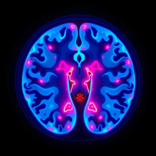
In recent advancements within neuropsychiatric imaging, a groundbreaking study has focused on a minute yet critical brain structure known as the habenula. This small but pivotal nucleus, nestled close to the thalamus, plays a vital regulatory role in monoaminergic signaling pathways, which are heavily implicated in mood regulation and depressive disorders. Despite its significance, the habenula’s tiny size and inherent variability among individuals have historically hindered consistent imaging analyses and the understanding of its alterations in depression.
The new research leverages cutting-edge high-resolution structural magnetic resonance imaging (MRI) at 3-Tesla strength to probe the habenula’s intricacies in patients experiencing their first episode of depression, who have not yet undergone any antidepressant treatment. This focus on first-episode depression (FED) patients eliminates confounding effects of long-term medication or illness chronicity, allowing for a purer insight into the neurobiological underpinnings of early depressive pathology.
Traditional MRI assessments often rely on gross volumetric analysis or signal intensity parameters, but such approaches may overlook subtle, spatially heterogeneous changes within the habenular complex. To circumvent this limitation, the study incorporated advanced voxel-based radiomic analysis coupled with clustering algorithms. Radiomics extracts a high-dimensional array of quantitative imaging features that capture voxel-level variations in texture, intensity, and spatial distribution, potentially revealing microstructural and molecular alterations invisible to conventional imaging metrics.
The investigation enrolled 94 participants split evenly between healthy controls and patients with first-episode depression. Precise segmentation of the habenula was performed, followed by detailed measurements of volumetric size and T1 relaxation times—a parameter sensitive to tissue composition and microenvironment. These neuroimaging markers were analyzed in relation to age, depression severity, and other clinical parameters, providing an integrative picture of habenular changes associated with early depressive states.
Interestingly, the study found a positive correlation between habenular T1 values and age in healthy controls, aligning with expected age-associated tissue modulation. However, this relationship was absent in patients with first-episode depression, suggesting that depression may disrupt normal age-related microstructural dynamics within this crucial brain region. Such findings underscore the habenula’s complex role as a neurobiological hub potentially vulnerable to pathological modification in mood disorders.
Beyond simple measures, the researchers applied a clustering-based radiomics model to classify participants into depression or control groups. This sophisticated model outperformed traditional imaging analyses, achieving an impressive area under the curve (AUC) of 0.844 compared to 0.708 for conventional approaches. This jump in diagnostic accuracy highlights the potential for machine learning-enhanced radiomic signatures of the habenula to serve as biomarkers for early depressive episodes.
The heterogeneity captured by the clustering method likely reflects nuanced microarchitectural changes within the habenula that conventional volume or mean signal assessments fail to detect. These subtle texture and intensity patterns may correspond to underlying pathophysiological processes—such as altered synaptic density, neurotransmitter receptor expression, or glial activity—thereby opening avenues for mechanistic and therapeutic exploration.
Crucially, this work emphasizes the habenula’s internal variability as not just an imaging curiosity but as a meaningful biological signal intimately tied to depression. By moving beyond simplistic gross anatomy to embrace radiomic complexity, the study bridges cutting-edge neuroimaging with clinical psychiatry, fostering innovative diagnostic and potentially prognostic tools.
The potential clinical implications are profound. Early, accurate identification of depression using noninvasive imaging biomarkers can revolutionize patient stratification, enabling personalized interventions at a critical illness juncture. Moreover, monitoring habenular alterations over time could serve as a surrogate marker for treatment response or illness progression, guiding clinicians towards more tailored management strategies.
This study also sparks curiosity about the habenula’s broader role in neuropsychiatric conditions characterized by monoaminergic dysregulation. Future research may expand these radiomic methodologies to bipolar disorder, schizophrenia, or treatment-resistant depression, further elucidating shared and unique neural circuit abnormalities.
Pioneering work like this underscores the transformative power of integrating advanced image processing, machine learning, and robust neurobiological theory. As functional and structural imaging reach ever finer resolutions, the ability to decode complex brain regions such as the habenula transforms from a technical challenge into a diagnostic opportunity.
By illustrating that the habenula’s internal heterogeneity carries diagnostic significance, the study pioneers a paradigm shift in psychiatric imaging. It challenges the field to reconsider simplistic volumetric analyses and embrace multivariate, data-driven approaches that capture the brain’s intricate microstructure in health and disease.
Ultimately, this research highlights the promise of precision psychiatry, where imaging biomarkers derived from radiomics and cluster analysis inform early, accurate diagnosis and illuminate pathophysiology. The habenula emerges as a compelling locus for such innovation, advancing our understanding of depression’s neural substrates and opening new vistas for intervention.
The study’s findings invite an optimistic future where fusion of neuroimaging technology, computational analytics, and clinical insight coalesce to redefine mental health diagnosis. Such advances could dramatically enhance early detection and targeted treatment, potentially mitigating the global burden of depression through improved neuroscience-informed care.
Subject of Research: The microstructural and radiomic analysis of the habenula in first-episode depression using high-resolution 3-T MRI.
Article Title: High-resolution structural magnetic resonance examination of the Habenula in patients with first-episode depression: an exploratory radiomics diagnostic value analysis based on cluster analysis.
Article References: Hou, L., Bian, B., Luan, S. et al. High-resolution structural magnetic resonance examination of the Habenula in patients with first-episode depression: an exploratory radiomics diagnostic value analysis based on cluster analysis. BMC Psychiatry 25, 896 (2025). https://doi.org/10.1186/s12888-025-07259-4
Image Credits: AI Generated
DOI: https://doi.org/10.1186/s12888-025-07259-4
