In a groundbreaking advancement poised to revolutionize musculoskeletal oncology, researchers have unveiled a novel radiomics-based approach employing magnetic resonance imaging (MRI) to accurately grade chondroid bone tumors. These tumors, which encompass enchondromas and both low-grade and higher-grade chondrosarcomas, present a significant diagnostic challenge due to overlapping imaging characteristics and subtle histopathological differences. Harnessing sophisticated image analysis techniques, this emerging methodology promises to refine diagnostic precision, effectively guiding clinical decisions and potentially improving patient outcomes.
Chondroid tumors originate from cartilaginous cells within the bone, with enchondromas being benign lesions and chondrosarcomas representing malignant transformations with varying degrees of aggressiveness. Distinguishing these tumor grades has traditionally relied on invasive biopsy procedures and histopathological evaluation, both of which carry inherent limitations including sampling errors and procedural risks. Consequently, non-invasive imaging biomarkers capable of discerning tumor grade hold immense clinical appeal.
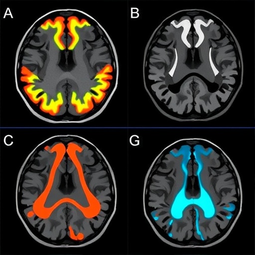
The research team embarked on a retrospective study involving 120 patients who underwent contrast-enhanced MRI examinations between 2009 and 2019. Their cohort included 92 cases of enchondromas, 16 low-grade chondrosarcomas, and 12 intermediate to high-grade chondrosarcomas, creating a robust dataset for analysis. Each tumor underwent meticulous manual segmentation by an expert musculoskeletal radiologist, with validation by a senior radiology consultant to ensure accuracy and consistency.
Central to this study was the application of radiomics—a cutting-edge analytic framework that extracts high-dimensional quantitative features from medical images beyond what the human eye can discern. These features capture subtle textural, shape, and intensity variations within tumor tissue, correlating with underlying pathophysiological processes. The researchers leveraged this data-rich environment to build predictive models capable of classifying tumors with high fidelity.
To optimize feature selection and classification, the study employed a two-pronged machine learning approach combining least absolute shrinkage and selection operator (LASSO) and random forest (RF) algorithms. LASSO served to reduce the dimensionality of extracted features by penalizing less informative variables, while random forest facilitated robust ensemble classification through decision tree aggregation. This synergy was designed to maximize predictive accuracy while mitigating overfitting risks inherent in high-dimensional data.
Recognizing the imbalance in tumor grade representation—particularly the comparatively fewer cases of higher-grade chondrosarcomas—the researchers incorporated the synthetic minority oversampling technique (SMOTE). SMOTE generates synthetic examples of minority class samples to balance the training dataset, preventing bias towards the more prevalent classes. Models were thus trained and tested both with and without SMOTE enhancement to assess its impact on classification performance.
Evaluation metrics focused on average precision, overall accuracy, area under the receiver operating characteristic curve (AUC), and weighted kappa statistics, providing a comprehensive assessment of model reliability. Notably, the combined LASSO plus random forest model trained on all MRI sequences outperformed others, achieving a striking accuracy of approximately 82.6% and an AUC nearing 0.97. These figures underscore the model’s exceptional ability to discriminate among tumor subtypes.
Interestingly, the model utilizing T2-weighted imaging sequences paired with SMOTE enhancement achieved the highest mean average precision (mAP) of 0.75, signaling the critical role that managing class imbalance plays in refining predictions. This finding aligns with broader machine learning literature emphasizing balanced datasets for optimal classifier training, especially in medical imaging contexts where pathological heterogeneity is common.
Quadratic weighted kappa values ranged from 0.65 to 0.73 across the evaluated models, which translates to substantial agreement when cross-referenced with pathological diagnoses. This statistic measures the concordance between predicted classification and ground truth, implying that the radiomics-driven approach closely approximates gold-standard histopathology without invasive procedures.
The implications of this work extend beyond mere diagnostic refinement. By providing clinicians with a non-invasive, highly accurate tool for tumor grading, patient management could be revolutionized through tailored treatment regimens. Accurate differentiation between benign and malignant chondroid lesions is paramount for determining the necessity of surgical intervention or conservative monitoring, directly impacting morbidity and healthcare resources.
Further, this radiomics framework suggests a path toward integrating artificial intelligence into routine musculoskeletal imaging workflows. As MRI is widely accessible and routinely employed in clinical practice, embedding these analytic techniques could enable real-time decision support, augmenting radiologist expertise and standardizing assessments across institutions.
While the study’s retrospective design and relatively modest sample size emphasize the need for prospective validation in larger, multi-center cohorts, its findings establish a compelling proof-of-concept. Expanding such research will be crucial to ascertain generalizability across different MRI platforms, scanning protocols, and patient demographics.
Moreover, future investigations may explore the fusion of radiomics features with other omics data—such as genomics or proteomics—to further enhance tumor characterization. The integration of multi-modal data promises a holistic understanding of tumor biology, ultimately driving personalized medicine approaches in orthopedic oncology.
This advancement indicates a paradigm shift in the evaluation of cartilaginous bone tumors, reducing dependence on invasive tissue sampling, and mitigating associated risks. Patients stand to benefit from quicker, less burdensome diagnoses and optimized therapeutic strategies tailored to the biological aggressiveness of their lesions.
In conclusion, the integration of MRI-based radiomics and advanced machine learning algorithms offers a potent solution to the longstanding challenge of grading chondroid tumors with high precision. This method’s ability to distinguish benign enchondromas from low- and high-grade chondrosarcomas non-invasively heralds a future where artificial intelligence augments clinical acumen in musculoskeletal oncology.
As research in this domain accelerates, we may soon witness widespread adoption of radiomics pipelines in radiology departments worldwide, transforming how bone tumors are diagnosed, graded, and managed. The cross-disciplinary collaboration between radiologists, oncologists, data scientists, and bioinformaticians reflects the innovative frontier pushing personalized healthcare.
Ultimately, this study exemplifies the promise of combining medical imaging with computational prowess to solve intricate clinical problems. With continued refinement and expansive validation, MRI radiomics stands to become an indispensable ally in the fight against bone cancers, improving diagnostic accuracy and patient care worldwide.
Subject of Research:
Development of a multiclass radiomics model utilizing preoperative MRI to differentiate between enchondroma, low-grade chondrosarcoma, and higher-grade chondrosarcoma.
Article Title:
Grading chondroid tumors through MRI radiomics: enchondroma, low-grade chondrosarcoma and higher-grade chondrosarcoma.
Article References:
Park, H., Lee, J., Lee, S. et al. Grading chondroid tumors through MRI radiomics: enchondroma, low-grade chondrosarcoma and higher-grade chondrosarcoma. BMC Cancer 25, 918 (2025). https://doi.org/10.1186/s12885-025-14330-6
Image Credits: Scienmag.com
